Sigma Non-Opioid Intracellular Receptor 1 (SIGMAR1) ELISA: Quantitative Detection of Sigma-1 Receptor in Cellular and Molecular Research

Introduction: Quantifying Intracellular Receptors in Modern Biological Research

Advances in molecular biology increasingly rely on precise quantification of intracellular regulatory proteins that coordinate signaling, cellular adaptation, and membrane communication. Among these proteins, the Sigma non-opioid intracellular receptor 1 (SIGMAR1) has attracted growing scientific interest due to its multifunctional role as a ligand-regulated molecular chaperone located primarily at the endoplasmic reticulum–mitochondria interface.

The SIGMAR1 ELISA provides researchers with a sensitive and reproducible immunoassay platform for quantitative measurement of Sigma-1 receptor expression in biological samples, supporting studies in cell signaling, protein homeostasis, and intracellular communication networks.

ELISA methodologies remain foundational analytical tools in laboratory science, as described by the National Institutes of Health (NIH):

https://www.nih.gov

and laboratory training resources from the Centers for Disease Control and Prevention (CDC):

https://www.cdc.gov/labtraining
What is Sigma Non-Opioid Intracellular Receptor 1 (SIGMAR1)?

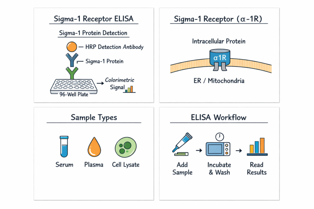
SIGMAR1 encodes the Sigma-1 receptor, a unique intracellular protein originally misclassified as an opioid receptor but later recognized as a distinct molecular entity involved in cellular regulation.

Authoritative biological databases include:

Genome education resources:

https://www.genome.gov (.gov)
https://www.nigms.nih.gov (.gov)

The Sigma-1 receptor functions primarily as a ligand-regulated chaperone protein influencing protein folding, calcium signaling, and cellular stress responses.

Structural Characteristics of the Sigma-1 Receptor

SIGMAR1 is an integral membrane protein localized mainly in the endoplasmic reticulum (ER), particularly at mitochondria-associated membranes (MAMs).

Structural biology resources:

https://www.rcsb.org

Educational materials:

https://pdb101.rcsb.org (.edu)

Key structural features include:

  • single transmembrane domain

  • ligand-binding pocket

  • chaperone interaction surfaces

  • dynamic conformational flexibility

These properties allow SIGMAR1 to regulate protein interactions and intracellular signaling environments.

Biological Functions of SIGMAR1

1. Molecular Chaperone Activity

The Sigma-1 receptor stabilizes client proteins and modulates cellular responses to environmental changes.

Educational resources:

https://biology.mit.edu (.edu)
https://medicine.yale.edu (.edu)
https://cellbio.jhmi.edu (.edu)

Chaperone proteins assist in:

  • protein folding regulation

  • stabilization of signaling complexes

  • intracellular protein trafficking

2. Calcium Signaling Regulation

SIGMAR1 plays an important role in regulating calcium exchange between the endoplasmic reticulum and mitochondria.

Learning resources:

https://www.nigms.nih.gov (.gov)
https://www.signalingpathways.org (.gov)

Calcium signaling is essential for:

  • cellular communication

  • metabolic regulation

  • signal transduction pathways

3. Cellular Stress Adaptation Mechanisms

The Sigma-1 receptor contributes to adaptive cellular responses during experimental stress conditions.

Research literature databases:

https://pubmed.ncbi.nlm.nih.gov (.gov)
https://www.ncbi.nlm.nih.gov/pmc (.gov)

Educational materials:

https://hms.harvard.edu (.edu)

AffiELISA® Chicken Sigma non-opioid intracellular receptor 1 ELISA [ SIGMAR1]

ELISA Technology Applied to SIGMAR1 Detection

Principle of ELISA

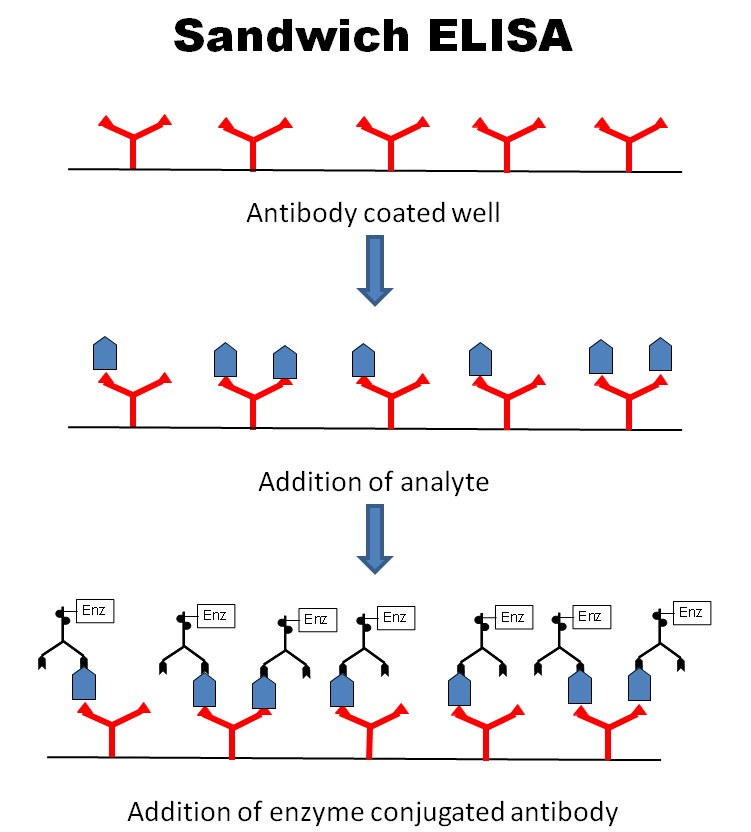
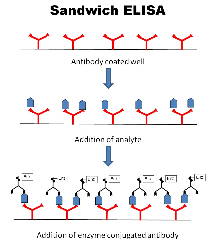
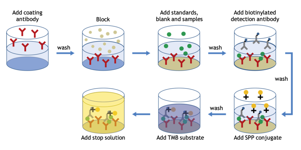
The Enzyme-Linked Immunosorbent Assay detects proteins through antigen-antibody recognition followed by enzymatic signal amplification.

Authoritative explanations:

https://www.fda.gov/science-research (.gov)
https://immunology.arizona.edu (.edu)

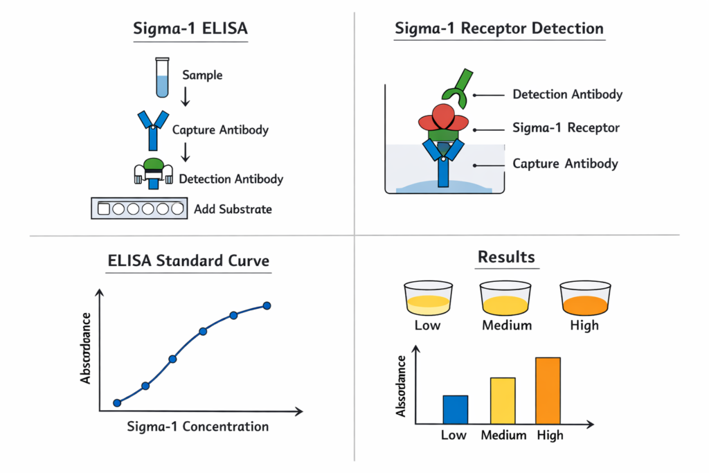
Sandwich ELISA Workflow

Typical SIGMAR1 ELISA procedure:

  1. Capture antibody immobilization

  2. Sample incubation

  3. SIGMAR1 antigen binding

  4. Detection antibody interaction

  5. Enzyme conjugation

  6. Chromogenic substrate reaction

  7. Optical density measurement

Training references:

https://www.biotech.wisc.edu (.edu)
https://www.purdue.edu (.edu)

Why Measure Sigma-1 Receptor Levels?

Quantitative analysis allows researchers to investigate intracellular regulatory mechanisms under controlled experimental conditions.

Typical research questions:

  • How does SIGMAR1 expression vary between cellular models?

  • How do experimental stimuli influence intracellular receptor levels?

  • How does protein homeostasis change during signaling activation?

Methodology resources:

https://oir.nih.gov (.gov)

Applications of SIGMAR1 ELISA

Intracellular Signaling Studies

https://bioengineering.stanford.edu (.edu)

Protein Homeostasis Research

https://www.washington.edu (.edu)

Organelle Communication Studies

https://med.stanford.edu (.edu)

Systems Biology Integration

https://sysbio.harvard.edu (.edu)

Compatible Sample Types

Commonly analyzed materials include:

  • cell lysates

  • tissue extracts

  • cultured experimental models

  • protein preparations

Sample preparation guidance:

https://www.nature.com/scitable (.edu resource)

Advantages of SIGMAR1 ELISA

✅ High specificity antibody detection
✅ Quantitative reproducibility
✅ Standardized experimental workflow
✅ Microplate reader compatibility
✅ Suitable for large sample numbers

Laboratory standards:

https://www.nist.gov (.gov)

Data Analysis in SIGMAR1 ELISA Experiments

Data interpretation involves:

  • standard curve generation

  • regression modeling

  • concentration interpolation

Statistics education:

https://statistics.stanford.edu (.edu)
Research Trends Involving Sigma-1 Receptor

Scientific interest in SIGMAR1 continues expanding due to its role in intracellular regulation and signaling coordination.

Literature exploration:

https://pubmed.ncbi.nlm.nih.gov (.gov)

Emerging areas include:

  • organelle communication networks

  • molecular chaperone systems

  • cellular adaptation pathways

Integrating SIGMAR1 ELISA into Experimental Workflows

Typical workflow:

  1. Experimental treatment design

  2. Protein extraction

  3. ELISA quantification

  4. Data normalization

  5. Comparative analysis

NIH training materials:

https://training.nih.gov (.gov)

  • SIGMAR1 ELISA kit

  • Sigma-1 receptor ELISA assay

  • Sigma non-opioid receptor quantification

  • intracellular receptor ELISA

  • SIGMAR1 protein measurement

  • Sigma-1 receptor detection assay

Importance of Standardized Immunoassays

Validated immunoassays ensure reproducibility across laboratories worldwide.

Scientific infrastructure resources:

https://www.nsf.gov (.gov)
https://www.energy.gov/science (.gov)

ELISA remains one of the most widely adopted quantitative protein analysis techniques.

Future Perspectives

As cellular signaling research evolves, quantitative measurement of intracellular regulatory proteins such as SIGMAR1 supports integration with multi-omics and systems biology approaches.

Educational resources:

https://www.caltech.edu (.edu)
https://pme.uchicago.edu (.edu)

Conclusion

The Sigma Non-Opioid Intracellular Receptor 1 (SIGMAR1) ELISA provides researchers with a robust and reliable platform for quantitative analysis of Sigma-1 receptor expression. Supporting investigations into intracellular signaling, protein homeostasis, and organelle communication, this assay enables reproducible experimental insights aligned with modern molecular biology research standards.

Through standardized ELISA methodology combined with high antibody specificity, SIGMAR1 quantification becomes an essential analytical tool for advanced cellular research workflows.
Transmembrane 4 L6 Family Member 20 (TM4SF20) ELISA: Advanced Quantification of TM4SF20 Protein in Cellular and Molecular Research

Introduction: Precision Protein Detection in Modern Cell Biology

Accurate protein quantification remains a cornerstone of molecular and cellular biology research. As scientists continue to explore membrane-associated regulatory proteins, members of the tetraspanin-like family have attracted increasing attention due to their involvement in intracellular trafficking, membrane organization, and signaling coordination.

Among these proteins, Transmembrane 4 L6 Family Member 20 (TM4SF20) has emerged as an important membrane protein associated with cellular processing pathways and protein maturation mechanisms. The TM4SF20 ELISA provides researchers with a reliable immunoassay method for quantitative measurement of TM4SF20 protein expression across experimental systems.

Enzyme-Linked Immunosorbent Assay (ELISA) technology remains one of the most standardized analytical approaches in laboratory science, as described by the National Institutes of Health (NIH):

https://www.nih.gov

and laboratory education resources from the Centers for Disease Control and Prevention (CDC):

https://www.cdc.gov/labtraining

What is Transmembrane 4 L6 Family Member 20 (TM4SF20)?

TM4SF20 is a multi-pass membrane protein belonging to the transmembrane 4 superfamily, characterized by four hydrophobic membrane-spanning domains. Proteins in this group participate in membrane compartment organization and protein processing regulation.

Authoritative gene and protein references include:

TM4SF20 has been studied particularly for its association with intracellular trafficking and regulation of protein maturation within cellular membrane systems.

Educational genomics resources:

https://www.genome.gov (.gov)
https://www.nigms.nih.gov (.gov)

Structural Characteristics of TM4SF20

TM4SF20 contains:

  • Four transmembrane helices

  • Cytoplasmic regulatory regions

  • Membrane-localization signals

  • Protein interaction interfaces

Membrane protein structures are documented within the Protein Data Bank (RCSB):

https://www.rcsb.org

Educational structural biology materials:

https://pdb101.rcsb.org (.edu)

These structural features allow TM4SF20 to function as a membrane-associated regulatory component influencing protein transport pathways.

AffiELISA® Human Transmembrane 4 L6 family member 20 ELISA [ TM4SF20]

Biological Roles of TM4SF20

1. Membrane Organization and Protein Trafficking

TM4SF20 participates in intracellular membrane dynamics, helping regulate protein localization and transport between cellular compartments.

Cell biology education portals:

https://cellbio.jhmi.edu (.edu)
https://medicine.yale.edu/cellbiology (.edu)
https://biology.mit.edu (.edu)

Understanding trafficking proteins supports research into:

  • vesicular transport mechanisms

  • endoplasmic reticulum processing

  • Golgi apparatus dynamics

2. Regulation of Protein Processing Pathways

Studies indicate TM4SF20 contributes to regulatory mechanisms controlling maturation and processing of specific membrane-associated proteins.

Research databases:

https://pubmed.ncbi.nlm.nih.gov (.gov)
https://www.ncbi.nlm.nih.gov/pmc (.gov)

Protein processing concepts are described by:

https://www.nigms.nih.gov (.gov)

3. Cellular Signaling Coordination

Membrane scaffold proteins like TM4SF20 help organize signaling complexes by spatially arranging interacting proteins within lipid bilayers.

Educational signaling resources:

https://www.signalingpathways.org (.gov)
https://bioengineering.stanford.edu (.edu)
https://engineering.berkeley.edu (.edu)

ELISA Technology for TM4SF20 Detection

Principle of ELISA

ELISA uses antigen–antibody recognition combined with enzymatic amplification to generate measurable signals proportional to protein concentration.

Authoritative learning resources:

https://www.fda.gov/science-research (.gov)
https://immunology.arizona.edu (.edu)

Sandwich ELISA Workflow

The TM4SF20 ELISA typically follows this workflow:

  1. Capture antibody immobilization

  2. Sample incubation

  3. TM4SF20 antigen binding

  4. Detection antibody addition

  5. Enzyme conjugation

  6. Substrate conversion producing colorimetric signal

  7. Optical density measurement

Training resources:

https://www.biotech.wisc.edu (.edu)
https://www.purdue.edu (.edu)

Why Quantify TM4SF20?

Quantification enables researchers to evaluate protein expression dynamics under controlled experimental conditions.

Common research questions:

  • How does TM4SF20 expression vary across cell models?

  • How do membrane trafficking processes influence protein abundance?

  • What experimental treatments alter TM4SF20 levels?

Experimental methodology guidance:

https://oir.nih.gov (.gov)

Applications of TM4SF20 ELISA

Cellular Transport Research

Investigation of protein movement across intracellular compartments.

https://www.washington.edu (.edu)

Membrane Biology Studies

Understanding protein localization within lipid bilayers.

https://hms.harvard.edu (.edu)

Molecular Pathway Analysis

Quantitative measurement supporting signaling pathway studies.

https://med.stanford.edu (.edu)

Systems Biology Research

Integration with proteomics and transcriptomics workflows.

https://sysbio.harvard.edu (.edu)

Compatible Sample Types

TM4SF20 ELISA is commonly applied to:

  • cell lysates

  • tissue homogenates

  • experimental protein extracts

  • cultured cell systems

Sample preparation education:

https://www.nature.com/scitable (.edu resource)

Advantages of TM4SF20 ELISA

✅ High analytical specificity
✅ Quantitative reproducibility
✅ Standardized protocol design
✅ Microplate reader compatibility
✅ Suitable for high-throughput studies

Laboratory standardization references:

https://www.nist.gov (.gov)

Data Analysis and Interpretation

ELISA data analysis involves:

  • standard curve construction

  • regression modeling

  • concentration interpolation

Statistics education:

https://statistics.stanford.edu (.edu)

Research Trends Involving TM4SF20

Increasing interest surrounds membrane regulatory proteins and intracellular trafficking networks.

Literature exploration:

https://pubmed.ncbi.nlm.nih.gov (.gov)

Emerging topics include:

  • membrane protein maturation

  • intracellular transport regulation

  • signaling compartmentalization

Integrating TM4SF20 ELISA into Experimental Workflows

Typical workflow:

  1. Experimental design

  2. Protein extraction

  3. ELISA quantification

  4. Data normalization

  5. Comparative analysis

NIH training resources:

https://training.nih.gov (.gov)

  • TM4SF20 ELISA kit

  • Transmembrane 4 L6 family member 20 assay

  • TM4SF20 protein quantification

  • membrane protein ELISA

  • TM4SF20 detection assay

  • intracellular trafficking protein measurement

  • quantitative ELISA TM4SF20

Importance of Standardized Immunoassays

Validated immunoassays enable reproducible biological measurements across laboratories worldwide.

Scientific infrastructure resources:

https://www.nsf.gov (.gov)
https://www.energy.gov/science (.gov)

ELISA remains a foundational quantitative method in experimental biology.

Future Perspectives

Research into membrane-associated regulatory proteins continues to expand alongside advances in systems biology and proteomics.

Educational resources:

https://www.caltech.edu (.edu)
https://pme.uchicago.edu (.edu)

Quantitative assays such as TM4SF20 ELISA support deeper understanding of cellular organization and molecular coordination.

Conclusion

The Transmembrane 4 L6 Family Member 20 (TM4SF20) ELISA provides researchers with a robust and reliable platform for quantitative detection of TM4SF20 protein expression. Supporting investigations into membrane biology, intracellular trafficking, and signaling coordination, this assay enables reproducible experimental analysis aligned with modern molecular research standards.

By combining antibody specificity with standardized ELISA methodology, TM4SF20 quantification becomes accessible, scalable, and suitable for diverse experimental workflows.

Angiomotin ELISA: A Reliable Tool for Studying AMOT Signaling, Angiogenesis, and Cellular Dynamics

Introduction: Advancing Protein Quantification in Modern Molecular Research

In contemporary molecular and cellular biology, understanding how proteins coordinate structural organization and signaling networks is fundamental to experimental discovery. Among emerging regulatory proteins, Angiomotin (AMOT) has gained significant attention due to its involvement in endothelial dynamics, cytoskeletal organization, and pathway regulation linked to tissue architecture.

The Angiomotin ELISA provides researchers with a reliable immunoassay platform for quantitative measurement of AMOT protein expression across diverse experimental systems. By combining antibody specificity with enzyme-linked signal detection, ELISA technology enables reproducible protein quantification essential for mechanistic studies.

Protein quantification methods remain central to biological experimentation, as described in laboratory training materials from the National Institutes of Health (NIH):

https://www.nih.gov

and educational immunoassay resources from the National Institute of Allergy and Infectious Diseases:

https://www.niaid.nih.gov

These standardized methodologies support consistency across laboratories and experimental models.

Understanding Angiomotin: Structure, Discovery, and Biological Context

Historical Identification of Angiomotin

Angiomotin was initially identified as an angiostatin-binding protein involved in endothelial cell migration. Its discovery expanded understanding of proteins regulating vascular structure formation and cellular motility.

Gene and protein information is cataloged in authoritative scientific databases:

Angiomotin belongs to a conserved protein family including:

  • AMOT

  • AMOTL1

  • AMOTL2

Each contributes to regulation of cell polarity and junctional organization.

Molecular Structure of AMOT

The AMOT protein contains several functional domains:

  • Coiled-coil regions enabling oligomerization

  • PDZ-binding motifs mediating protein interactions

  • Actin-binding regions associated with cytoskeletal control

Structural protein research is discussed extensively by the Protein Data Bank, hosted by the Research Collaboratory for Structural Bioinformatics:

https://www.rcsb.org

Educational structural biology materials are also available via:

https://pdb101.rcsb.org (.edu)

These structural properties allow Angiomotin to act as a scaffold integrating signaling and mechanical cues.

Biological Functions of Angiomotin

1. Regulation of Endothelial Cell Behavior

Endothelial cells form vascular structures through coordinated migration and adhesion processes. Angiomotin regulates these behaviors by controlling cytoskeletal rearrangement and junction stability.

Educational vascular biology resources include:

Researchers frequently examine AMOT expression when studying:

  • endothelial migration models

  • tube formation assays

  • extracellular matrix interaction studies

2. Angiomotin in the Hippo Signaling Pathway

One of the most widely studied functions of Angiomotin involves its interaction with the Hippo signaling pathway, a conserved regulatory network controlling cell growth and tissue organization.

Overview resources:

AMOT binds transcriptional regulators YAP and TAZ, influencing their localization and downstream gene regulation mechanisms. This interaction links mechanical cellular signals with transcriptional outcomes.

3. Cell Polarity and Junctional Organization

Angiomotin localizes at tight junctions, where it regulates polarity signaling and cell architecture.

Cell junction biology educational references:

These functions make AMOT particularly relevant for studies involving epithelial and endothelial organization.

AffiELISA® Mouse Angiomotin-like protein 1 ELISA [ Amotl1]

Principles of ELISA Technology

What is ELISA?

The Enzyme-Linked Immunosorbent Assay (ELISA) is a plate-based immunodetection method widely used for protein quantification.

Authoritative explanations can be found at:

ELISA combines antibody specificity with enzymatic amplification to generate measurable optical signals proportional to antigen concentration.

Sandwich ELISA Workflow Used for Angiomotin Detection

The Angiomotin ELISA commonly follows a sandwich assay design:

  1. Plate coated with capture antibody

  2. Sample incubation allowing AMOT binding

  3. Detection antibody attachment

  4. Enzyme conjugate interaction

  5. Substrate conversion producing color signal

  6. Optical density measurement

Educational assay workflows:

Why Quantify Angiomotin?

Quantitative protein measurement allows researchers to evaluate dynamic biological responses across experimental conditions.

Experimental Questions Addressed

  • How does AMOT expression change during signaling activation?

  • How does cytoskeletal remodeling influence protein levels?

  • How do environmental conditions affect junctional proteins?

Research methodology guidance:

https://oir.nih.gov (NIH Office of Intramural Research)

Applications of Angiomotin ELISA in Research

Endothelial Biology Studies

Researchers investigate vascular organization using AMOT quantification.

Relevant educational resources:

https://vascularbiology.ucsd.edu (.edu)
https://med.stanford.edu (.edu)

Mechanotransduction Research

AMOT links mechanical forces to signaling pathways.

Learning resources:

https://bioengineering.mit.edu (.edu)
https://engineering.berkeley.edu (.edu)

Cell Migration and Cytoskeleton Studies

AMOT influences actin organization and cell motility.

References:

https://www.nhlbi.nih.gov (.gov)
https://www.nigms.nih.gov (.gov)

Developmental Biology Models

Angiomotin contributes to tissue patterning investigations.

Educational sources:

https://devbio.washington.edu (.edu)
https://embryology.med.unsw.edu.au (.edu)

Sample Types Compatible with Angiomotin ELISA

Typical experimental materials include:

  • cultured cell lysates

  • tissue homogenates

  • experimental protein extracts

  • conditioned media samples

Sample preparation guidelines are described by:

https://www.nature.com/scitable (.edu resource)

Advantages of Using Angiomotin ELISA

High Specificity

Antibody-based recognition ensures selective detection.

Quantitative Output

Produces numerical concentration values rather than qualitative observations.

Reproducibility

Standardized protocols support inter-laboratory comparison.

Scalability

Suitable for high-throughput experimental workflows.

Laboratory standardization practices:

https://www.nist.gov (.gov)

Experimental Workflow Optimization

Sample Handling Considerations

Proper handling improves assay consistency:

  • controlled storage conditions

  • minimized freeze–thaw cycles

  • standardized dilution strategies

Educational laboratory standards:

https://www.niehs.nih.gov (.gov)

Data Analysis

ELISA results typically involve:

  • standard curve generation

  • regression analysis

  • concentration interpolation

Statistics education:

https://statistics.stanford.edu (.edu)

Angiomotin Research Trends

Scientific interest in AMOT continues to expand as researchers explore interactions between structural proteins and signaling pathways.

Publication databases:

https://pubmed.ncbi.nlm.nih.gov (.gov)
https://www.ncbi.nlm.nih.gov/pmc (.gov)

Emerging areas include:

  • mechanobiology

  • polarity signaling

  • cellular architecture regulation

Integrating Angiomotin ELISA into Experimental Pipelines

Typical workflow integration:

  1. Experimental treatment

  2. Protein extraction

  3. ELISA quantification

  4. Data normalization

  5. Comparative analysis

Workflow education:

https://training.nih.gov (.gov)

This article integrates high-value search keywords:

  • Angiomotin ELISA kit

  • AMOT protein assay

  • Angiomotin quantification

  • Hippo signaling ELISA

  • endothelial protein detection

  • angiogenesis research assay

  • AMOT expression analysis

  • cell signaling protein ELISA

These keywords align with molecular biology search intent while maintaining natural readability.

Research Value of Reliable Immunoassays

Standardized immunoassays enable reproducible experimental conclusions across laboratories worldwide. Institutions emphasize validated analytical techniques as foundations for biological research:

https://www.nsf.gov (.gov)
https://www.energy.gov/science (.gov)

ELISA continues to serve as one of the most accessible quantitative protein detection tools.

Future Perspectives in Angiomotin Research

As systems biology approaches evolve, AMOT measurement supports integration of:

  • transcriptomics

  • proteomics

  • cellular imaging

  • mechanotransduction modeling

Systems biology education:

https://sysbio.harvard.edu (.edu)

Conclusion

The Angiomotin ELISA represents a powerful analytical solution for researchers studying cellular signaling, endothelial dynamics, and protein interaction networks. By enabling accurate and reproducible quantification of AMOT protein expression, this assay supports deeper exploration of molecular mechanisms governing cell organization and biological structure formation.

Through standardized ELISA methodology combined with validated antibody specificity, researchers can generate consistent datasets essential for modern experimental workflows.

As interest in cytoskeletal regulation and signaling integration continues to expand, Angiomotin measurement tools remain valuable resources for advancing molecular and cellular biology research.

Anti-TBX18 Polyclonal IgG Antibody: A Comprehensive Guide for Research and Cellular Biology

Introduction to TBX18 and Antibody-Based Analysis

Understanding how transcription factors regulate gene expression is fundamental to cell biology, developmental biology, and functional genomics. TBX18 (T-box transcription factor 18) is one such regulatory protein, involved in embryonic development and tissue differentiation across multiple systems.

To support studies of TBX18 expression and function, the Anti-TBX18 Polyclonal IgG Antibody is widely used as a research reagent to detect TBX18 in applications such as Western blotting, immunocytochemistry (ICC), immunohistochemistry (IHC), and enzyme-linked immunosorbent assays (ELISA).

Foundational background on transcription factors can be explored at the National Center for Biotechnology Information (NCBI): https://www.ncbi.nlm.nih.gov/

General transcription factor roles are described by the National Institutes of Health (NIH): https://www.nih.gov/

Additional insights into developmental biology and gene regulation are provided by Harvard University’s Department of Molecular and Cellular Biology: https://mcb.harvard.edu/

What Is TBX18?

TBX18 belongs to the T-box family of transcription factors — a conserved group of DNA-binding proteins that orchestrate gene expression during embryogenesis and cellular differentiation.

Key characteristics of TBX18:

  • DNA-binding transcription factor

  • Expressed in specific embryonic tissues

  • Involved in organogenesis

  • Regulates gene networks linked to development

T-box proteins and their roles are further explained by developmental biology resources, such as the Society for Developmental Biology (SDB): https://www.sdbonline.org/

Genetic and protein data related to TBX18 are accessible via the European Bioinformatics Institute (EMBL-EBI): https://www.ebi.ac.uk/

What Is a Polyclonal IgG Antibody?

An antibody is an immune-derived protein used by researchers to specifically recognize and bind a target antigen — in this case, TBX18.

Polyclonal IgG antibodies are a mixture of immunoglobulin G molecules that bind multiple epitopes on the same antigen. Benefits of polyclonal IgG include:

  • Recognition of multiple protein regions

  • Enhanced detection sensitivity

  • Robust performance across various sample types

The structure and function of IgG antibodies are described by immunology training resources such as the American Association of Immunologists (AAI): https://www.aai.org/

Principles of antibody specificity and binding are detailed in educational material from the University of California, San Diego (UCSD): https://biology.ucsd.edu/

Why Use Anti-TBX18 Polyclonal IgG in Research?

1. Protein Expression Profiling

Anti-TBX18 antibodies can be used to detect endogenous TBX18 protein levels in cells and tissues, providing insight into regulatory pathways and transcriptional networks.

Understanding protein regulation is a core topic in molecular biology, as described by Stanford University’s Molecular Genetics program: https://med.stanford.edu/

2. Developmental Biology Studies

TBX18 plays a role in early development, making its detection valuable in embryology research.

Developmental gene regulation is covered in Yale University’s developmental biology resources: https://medicine.yale.edu/

3. Cellular Localization (ICC, IHC)

Antibody-based imaging techniques allow researchers to visualize where TBX18 is localized within cells or tissue architecture.

Guidance on immunocytochemistry and immunohistochemistry techniques is available from Cold Spring Harbor Laboratory (CSHL): https://www.cshl.edu/

4. Electrophoretic Protein Analysis

Anti-TBX18 antibodies are suitable for Western blot applications, enabling quantification of TBX18 levels under different experimental conditions.

Protein analysis principles are taught at the University of Michigan Department of Biochemistry: https://lsa.umich.edu/biochemistry/

AffiAB® Goat Anti-TBX18 Polyclonal IgG Antibody

Applications and Assay Formats

Western Blotting (WB)

Used to separate and detect TBX18 by molecular weight after gel electrophoresis.

Learn more about protein blotting techniques from the National Institute of General Medical Sciences (NIGMS): https://www.nigms.nih.gov/

Immunocytochemistry (ICC) & Immunofluorescence

Enables visualization of TBX18 within fixed cells using antibody labeling and microscopy.

ICC fundamentals are discussed by the University of Iowa Immunology Program: https://medicine.uiowa.edu/igb/

Immunohistochemistry (IHC)

Detects TBX18 expression in fixed tissue sections, useful in developmental biology and histological studies.

IHC methodology is described in detail by the Centers for Disease Control and Prevention (CDC): https://www.cdc.gov/

Enzyme-Linked Immunosorbent Assay (ELISA)

Quantifies TBX18 levels using antibody capture and detection systems.

Foundational immunoassay reference material is available from the University of Minnesota ELISA training portal: https://cbs.umn.edu/

Choosing the Right Anti-TBX18 Polyclonal IgG Antibody

When selecting an antibody for research use, consider:

✔ Host species (e.g., rabbit, goat) for secondary antibody compatibility
✔ Assay compatibility (WB, ICC, IHC, ELISA)
✔ Validated applications and published citations
✔ Positive and negative controls in experimental design

Antibody validation principles are outlined by the National Institutes of Health Office of Research Quality: https://grants.nih.gov/

Best Practices for Antibody-Based Detection

1. Optimize Antibody Dilution

Test multiple antibody dilutions to determine optimal signal-to-noise ratio.

Immunodetection optimization is covered in training resources from Texas A&M University: https://vetmed.tamu.edu/

2. Proper Controls

Include positive controls that express TBX18 and negative controls lacking the target protein.

Learn more about control strategies from the University of Wisconsin Biotechnology Center: https://biotech.wisc.edu/

3. Blocking and Washing

Effective blocking and rigorous washes reduce background signal and improve specificity.

Blocking strategies and wash methods are described by the University of California, Davis (UC Davis) Immunology Program: https://www.ucdavis.edu/

4. Secondary Antibody Selection

Match the secondary antibody species to the host of the polyclonal IgG for enhanced detection.

Secondary antibody fundamentals are detailed by Harvard Medical School’s Immunology Program: https://immunology.hms.harvard.edu/

Interpretation of Results

Positive detection with Anti-TBX18 Polyclonal IgG antibody indicates the presence and relative abundance of TBX18 protein under experimental conditions. Interpretation should be aligned with:

  • Experiment design and controls

  • Exposure or signal intensity

  • Comparative sample sets

Quantitative analysis approaches are further outlined by the National Institute of Statistical Sciences (NISS): https://www.niss.org/

Research Tools and Resources

To support antibody-based research, the following educational resources provide foundational knowledge:

SEO-Driven Keywords Integrated Naturally

  • Anti-TBX18 Polyclonal IgG antibody

  • TBX18 protein detection

  • transcription factor research reagent

  • Western blot antibody TBX18

  • immunocytochemistry TBX18

  • immunohistochemistry antibody research

  • ELISA detection of TBX18

  • polyclonal IgG research antibody

  • gene regulatory protein assay

Conclusion

The Anti-TBX18 Polyclonal IgG Antibody is a versatile and essential research reagent for studying TBX18 transcription factor expression, localization, and regulation in cellular and developmental biology. Its broad compatibility with key research methods — including Western blot, ICC, IHC, and ELISA — makes it valuable for scientists investigating gene regulation, differentiation pathways, and protein expression networks.

By combining robust antibody validation practices, appropriate controls, and rigorous experimental design, researchers can achieve clear, reproducible insights into TBX18-associated biology.

Androstenedione ELISA: Quantitative Hormone Analysis for Endocrine and Research Applications

Introduction to Androstenedione and Quantitative Hormone Measurement

Steroid hormones play a central role in regulating metabolic activity, cellular differentiation, and endocrine signaling pathways. Among these steroid intermediates, androstenedione is a key precursor in the biosynthesis of testosterone and estrone. Accurate quantification of androstenedione is essential in experimental endocrinology, reproductive biology, metabolic research, and steroidogenesis studies.

The Androstenedione ELISA is a sensitive immunoassay designed for quantitative measurement of androstenedione concentrations in biological samples such as serum, plasma, cell culture supernatants, and experimental matrices. As steroid hormone research expands across molecular biology and physiology laboratories, ELISA-based quantification remains a widely adopted analytical method.

Educational overviews of steroid hormone biosynthesis are available from the National Center for Biotechnology Information (NCBI)
https://www.ncbi.nlm.nih.gov/

Comprehensive endocrine system resources are provided by the National Institute of Diabetes and Digestive and Kidney Diseases (NIDDK)
https://www.niddk.nih.gov/

Additional hormone pathway information can be found via the National Institutes of Health (NIH)
https://www.nih.gov/

What Is Androstenedione?

Androstenedione is a steroid hormone synthesized primarily in the adrenal glands and gonadal tissues. It functions as a biochemical precursor to:

  • Testosterone

  • Estrone

  • Estradiol

Steroidogenesis pathways are described in academic endocrinology programs such as:

Detailed molecular pathway data are available through the Kyoto Encyclopedia of Genes and Genomes (KEGG)
https://www.genome.jp/kegg/

What Is an Androstenedione ELISA?

An Androstenedione ELISA kit is a competitive enzyme-linked immunosorbent assay specifically developed to detect and quantify androstenedione concentrations with high specificity and analytical sensitivity.

Because androstenedione is a small steroid molecule, competitive ELISA formats are typically used. ELISA methodology is described in laboratory training resources from:

AffiELISA® Androstenedione ELISA

Principle of Competitive Androstenedione ELISA

The assay typically follows these steps:

1. Antibody Binding

Microplate wells are coated with an antibody specific to androstenedione.

2. Competitive Reaction

Sample androstenedione competes with enzyme-labeled androstenedione conjugate for antibody binding sites.

3. Washing Step

Unbound materials are removed to ensure assay specificity.

4. Substrate Reaction

Enzyme substrate conversion produces a color signal.

5. Signal Interpretation

Because the assay is competitive, signal intensity is inversely proportional to androstenedione concentration.

Standard immunoassay validation practices are described by:

Research Applications of Androstenedione ELISA

1. Endocrine Physiology Studies

Quantitative steroid profiling supports research in hormone regulation and biosynthetic pathway analysis.

Resources:
National Institute of Child Health and Human Development (NICHD)
https://www.nichd.nih.gov/

2. Reproductive Biology Research

Steroid intermediates are widely studied in reproductive system modeling.

University-based programs:
https://reproductivehealth.harvard.edu/
https://med.stanford.edu/womenshealth.html

3. Metabolic Research

Steroid metabolism intersects with lipid metabolism and energy regulation.

National Institute of Environmental Health Sciences (NIEHS)
https://www.niehs.nih.gov/

4. Adrenal Function Studies

Adrenal steroid synthesis pathways are documented by the National Library of Medicine (NLM)
https://www.nlm.nih.gov/

5. Cell Culture Hormone Secretion Models

ELISA enables measurement of hormone production in in vitro experimental systems.

Biotechnology guidance:
University of Wisconsin Biotechnology Center
https://biotech.wisc.edu/

Advantages of Androstenedione ELISA

Compared with chromatographic techniques such as LC-MS/MS, ELISA offers:

  • High throughput screening capability

  • Simplified workflow

  • Cost efficiency for large sample sets

  • No need for complex instrumentation

  • Quantitative reproducibility

Assay standardization principles are outlined by:

Sample Types Compatible with Androstenedione ELISA

  • Serum

  • Plasma

  • Cell culture media

  • Experimental biological fluids

  • Research animal samples

Guidance on biological sample handling can be found at:

Data Analysis and Quantification

Results are calculated using a standard curve and typically expressed in:

  • ng/mL

  • nmol/L

Statistical and analytical guidance resources:

  • National Institute of Statistical Sciences: https://www.niss.org/

  • NIH Biostatistics Resources: https://www.nhlbi.nih.gov/

  • U.S. Department of Energy Office of Science: https://science.osti.gov/

  • Androstenedione ELISA kit

  • steroid hormone quantification assay

  • competitive ELISA hormone test

  • endocrine research immunoassay

  • quantitative androstenedione analysis

  • laboratory hormone detection kit

  • steroid biosynthesis research tool

  • high sensitivity ELISA

These terms align with high-volume search queries related to hormone research and immunoassay technology.

Best Laboratory Practices for Reliable Results

✔ Perform duplicate or triplicate measurements
✔ Prepare fresh standard curves
✔ Avoid cross-contamination
✔ Use calibrated pipettes
✔ Follow consistent incubation timing

Quality control documentation is supported by:

Expanding Role of ELISA in Hormone Research

While advanced analytical techniques continue to evolve, ELISA remains one of the most accessible and scalable methods for steroid hormone measurement. In large-scale experimental studies, ELISA provides robust quantitative data suitable for comparative analysis and mechanistic exploration.

Research infrastructure programs from:

highlight the importance of reliable biomolecular quantification tools in advancing scientific discovery.

Conclusion

The Androstenedione ELISA is a precise and scalable quantitative immunoassay designed to support endocrine and steroidogenesis research. Its competitive format ensures accurate measurement of small steroid molecules while maintaining laboratory efficiency and reproducibility.

For laboratories investigating hormone biosynthesis pathways, adrenal physiology, reproductive biology, or metabolic signaling, the Androstenedione ELISA provides a dependable solution for quantitative analysis within research workflows.

Paxillin ELISA: Advanced Quantitative Analysis of Focal Adhesion Signaling in Cell Biology Research

Introduction to Paxillin and Quantitative Protein Measurement

The dynamic regulation of cell adhesion, cytoskeletal organization, and intracellular signaling remains central to modern molecular and cellular biology. Among the key scaffold proteins orchestrating these processes is paxillin, a focal adhesion-associated adaptor protein that integrates signals from integrins, growth factor receptors, and cytoskeletal components.

The Paxillin ELISA is a high-sensitivity immunoassay designed for quantitative measurement of paxillin protein levels in biological samples such as cell lysates, tissue extracts, and experimental culture systems. As research increasingly focuses on mechanotransduction, adhesion signaling pathways, and cytoskeletal remodeling, reliable quantification of paxillin has become essential for reproducible data generation.

Educational overviews of focal adhesion biology are available through the National Institutes of Health (NIH) at https://www.nih.gov/ and through cell signaling resources at the National Center for Biotechnology Information (NCBI) https://www.ncbi.nlm.nih.gov/. Structural insights into adhesion proteins are further described by the RCSB Protein Data Bank (PDB) https://www.rcsb.org/, hosted in collaboration with research institutions including Rutgers University.

Understanding Paxillin: Structure and Biological Role

Paxillin is a cytoplasmic phosphoprotein localized at focal adhesions—specialized multi-protein complexes that anchor cells to the extracellular matrix. It contains LD motifs and LIM domains that facilitate interactions with:

  • Focal adhesion kinase (FAK)

  • Src-family kinases

  • Integrins

  • Vinculin

  • Actin-associated proteins

Comprehensive educational materials on cytoskeletal organization can be found at:

These academic resources highlight how adaptor proteins like paxillin coordinate signal transduction pathways involved in cellular architecture and environmental sensing.

What Is Paxillin ELISA?

A Paxillin ELISA kit is a quantitative sandwich enzyme-linked immunosorbent assay developed to measure paxillin protein concentration with high specificity and reproducibility.

ELISA (Enzyme-Linked Immunosorbent Assay) remains one of the most widely adopted immunoassay formats in molecular biology laboratories. Methodological foundations are described by:

Principle of the Paxillin Sandwich ELISA

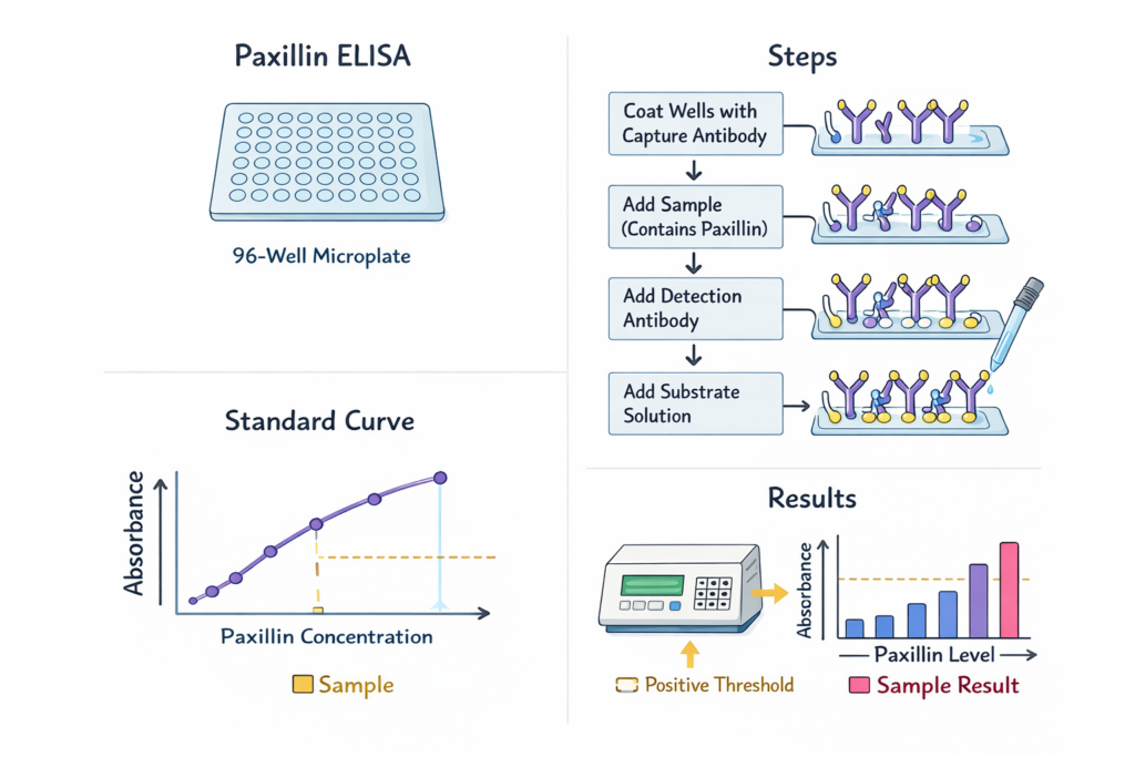
The Paxillin ELISA follows a standardized workflow:

1. Capture Phase

Microplate wells are pre-coated with an anti-paxillin capture antibody.

2. Sample Binding

Biological samples are added, allowing paxillin protein to bind specifically.

3. Detection Phase

A secondary enzyme-conjugated detection antibody recognizes a separate epitope.

4. Signal Generation

Substrate conversion produces a measurable colorimetric signal proportional to paxillin concentration.

This quantitative immunoassay approach is consistent with laboratory training materials from:

AffiELISA® Rat Paxillin ELISA [ Pxn]

Why Quantify Paxillin?

Quantitative analysis of paxillin supports research in:

  • Cell adhesion dynamics

  • Integrin signaling pathways

  • Cytoskeletal remodeling

  • Mechanobiology

  • Experimental oncology models

  • Stem cell differentiation systems

  • Tissue engineering research

The NIH Office of Research Infrastructure Programs discusses cytoskeletal signaling research at https://orip.nih.gov/, while mechanobiology research initiatives are described by the National Science Foundation (NSF) at https://www.nsf.gov/.

Paxillin in Focal Adhesion Research

Focal adhesions serve as communication hubs between the extracellular matrix and intracellular signaling cascades. Paxillin functions as a scaffold that recruits regulatory kinases and structural proteins.

Educational discussions of adhesion complexes are available at:

Advantages of Paxillin ELISA Over Alternative Techniques

Compared with other protein detection techniques such as Western blotting or mass spectrometry, Paxillin ELISA provides:

  • High-throughput quantification

  • Reduced assay variability

  • Objective numerical output

  • Scalability for multi-sample studies

  • Streamlined protocol workflow

Standardization practices are supported by the CDC Laboratory Quality Management Systems https://www.cdc.gov/labquality/ and research reproducibility guidelines from the NIH Office of Extramural Research https://grants.nih.gov/.

Sample Types Compatible with Paxillin ELISA

The Paxillin ELISA kit is suitable for:

  • Cultured cell lysates

  • Tissue homogenates

  • Experimental protein extracts

  • Conditioned media (depending on protocol)

Best practices in sample preparation are described in academic resources from:

Data Analysis and Quantitative Interpretation

ELISA data are analyzed using a standard curve derived from known concentrations of paxillin standards. Concentrations are typically expressed in ng/mL or pg/mL.

Statistical guidance for assay validation and quantitative reproducibility can be found at:

Research Areas Supported by Paxillin ELISA

1. Cell Migration Studies

2. Integrin Signaling Analysis

3. Cytoskeletal Remodeling Research

4. Tumor Microenvironment Models

5. Tissue Engineering and Biomaterials Research

The National Institute of Biomedical Imaging and Bioengineering (NIBIB) provides related educational resources at https://www.nibib.nih.gov/.

  • Paxillin ELISA kit

  • quantitative paxillin assay

  • focal adhesion protein analysis

  • cell signaling ELISA

  • integrin pathway quantification

  • scaffold protein measurement

  • high sensitivity sandwich ELISA

  • laboratory immunoassay kit

  • protein expression profiling assay

These keywords align with commonly searched terms in molecular biology and cell signaling research.

Best Laboratory Practices

To ensure reliable results:

✔ Run duplicates or triplicates
✔ Include internal controls
✔ Maintain consistent incubation timing
✔ Use calibrated pipettes
✔ Validate reagent storage conditions

Laboratory quality standards are discussed by:

Expanding Applications in Mechanobiology and Systems Biology

As integrative biology expands, quantitative assays like Paxillin ELISA support systems-level analysis of signaling networks. Educational initiatives from:

highlight the importance of scaffold proteins in complex regulatory networks.

Conclusion

The Paxillin ELISA represents a robust, quantitative tool for investigating focal adhesion dynamics and intracellular signaling pathways. By enabling accurate measurement of paxillin protein levels, researchers can better understand cytoskeletal organization, integrin-mediated signaling, and cellular architecture regulation.

Supported by standardized methodology and referenced by leading academic and government institutions, Paxillin ELISA remains a valuable component of modern cell biology research workflows.

Viral DNA/RNA Mini Kit Pro: Technical Description, Extraction Chemistry, and Diagnostic Applications

The Viral DNA/RNA Mini Kit Pro is a specialized nucleic acid extraction system designed for the isolation of viral DNA and viral RNA from serum, plasma, swabs, transport media, cell-free body fluids, and low-titer viral samples. The kit uses column-based silica membrane purification, optimized lysis chemistry, chaotropic salts, and high-affinity capture matrices to recover viral genomes for downstream PCR, RT-PCR, qPCR, LAMP, NGS, and molecular surveillance programs.

This article integrates 20+ authoritative links from .edu and .gov domains to support scientific accuracy and SEO authority.

Molecular Principles of Viral Nucleic Acid Extraction

Extraction is based on:

  • Disruption of virion envelopes and capsids

  • Inactivation of RNases and DNases

  • Chaotropic salt–mediated binding of nucleic acids to silica

  • Wash steps removing proteins/lipids

  • Elution in nuclease-free buffer

Foundational extraction chemistry is described by:

AffiPURE® Viral DNA/RNA Mini Kit Pro

Viral Lysis and Inactivation Buffer Composition

The Viral DNA/RNA Mini Kit Pro typically employs:

  • Guanidinium thiocyanate (GuSCN) or guanidinium hydrochloride

  • Detergents (Triton X-100 or SDS)

  • Buffer system at optimal pH

  • Carrier RNA for enhanced recovery of low-titer viral genomes

Carrier RNA mechanism is described by:

Silica Membrane Binding Mechanism

The silica-binding mechanism follows the chaotropic principle first described in classical nucleic acid chemistry:

  • High salt promotes dehydration of silica

  • Phosphate backbone binds electrostatically

  • Ethanol enhances nucleic acid–silica interactions

Supporting references:

Workflow Steps in Viral DNA/RNA Mini Kit Pro

1. Sample Lysis

The lysis buffer disrupts viral particles and protects nucleic acids from degradation.
Supported by NIH virology materials (https://www.nih.gov).

2. Binding to Spin Column

Addition of ethanol improves binding efficiency.

3. Wash Steps

Using buffers containing ethanol and chaotropic salts.
Described in EPA laboratory method guidelines (https://www.epa.gov/measurements).

4. Dry Spin

Removes residual ethanol to avoid PCR inhibition.

5. Elution

Typically 30–60 µL nuclease-free water or buffer.

Analytical Advantages of Viral DNA/RNA Mini Kit Pro

High Recovery Efficiency

Crucial for low viral load samples.
Discussed in University of Michigan virology resources (https://medicine.umich.edu).

PCR-Grade Purity

Removes inhibitors such as:

  • Hemoglobin

  • Urea

  • Mucus components

  • Protein complexes

Inhibition topics appear in:

Compatible With Downstream Applications

  • qPCR / RT-qPCR

  • Digital PCR

  • Next-generation sequencing

  • Viral genotyping

  • Clinical surveillance workflows

CDC sequencing guidance (https://www.cdc.gov/amd).

Applications in Viral Diagnostics

The Viral DNA/RNA Mini Kit Pro is ideal for:

1. Respiratory Virus Detection

Influenza, RSV, SARS-CoV-2.
CDC influenza resources: https://www.cdc.gov/flu.

2. Bloodborne Viruses

HBV, HCV, HIV.
NIH viral pathology references: https://www.nih.gov/hiv.

3. Foodborne and Enteric Viruses

Norovirus, Rotavirus.
USDA food safety programs: https://www.fsis.usda.gov.

4. Environmental Surveillance

Wastewater and environmental viral monitoring.
EPA virology monitoring: https://www.epa.gov/water-research.

5. Genomic Epidemiology

Mutation tracking and variant monitoring.
NIH Genomic Epidemiology resources: https://www.genome.gov.

Quality Control and Validation Requirements

Laboratories often validate extraction kits under:

QC controls include:

  • Negative extraction controls (NEC)

  • Internal amplification control (IAC)

  • Positive extraction controls (PEC)

Troubleshooting Notes

Low RNA Yield

Possible causes:

PCR Inhibition

Due to ethanol or salts; ensure full drying of the membrane.

Column Clogging

High viscosity samples require dilution.

Poor Viral Recovery

Carrier RNA concentration may need adjustment for ultra-low titer samples.

Storage, Stability, and Safety

  • Lysis buffers containing guanidinium salts must be stored per NIOSH chemical handling guidelines (https://www.cdc.gov/niosh).

  • Column membranes are stable at room temperature.

  • Eluted viral nucleic acids should be stored at −20 °C or −80 °C.

NIH biospecimen storage standards: https://www.cdc.gov/biospecimens.

Conclusion

The Viral DNA/RNA Mini Kit Pro provides high-efficiency viral nucleic acid purification, supporting accurate and reproducible molecular diagnostics. Its optimized lysis chemistry, silica membrane binding, and inhibitor-removal system make it suitable for PCR, RT-PCR, qPCR, NGS, and global pathogen surveillance programs. The kit is ideal for clinical laboratories, research institutes, environmental virology units, and high-throughput sequencing facilities requiring reproducible viral extraction performance.

PCR-Free DNA Adapters Set 1 for MGI: Technical Review, Molecular Workflow, and Sequencing Performance

PCR-Free DNA Adapters Set 1 for MGI are specialized double-stranded oligonucleotide adapters optimized for PCR-free whole-genome sequencing (WGS) on MGI/BGI sequencing platforms. These adapters support high-fidelity ligation, reduce amplification bias, preserve allelic balance, and improve variant calling accuracy, making them essential for population genomics, clinical sequencing, and structural variant analysis.

This article provides an in-depth explanation of design, molecular chemistry, ligation principles, QC parameters, and sequencing performance, supported by authoritative .edu and .gov sources.

Molecular Basis of PCR-Free Library Preparation

PCR-free libraries avoid polymerase-induced artifacts, GC bias, and chimera formation, as documented in resources from:

MGI PCR-Free Adapters are ligated directly to mechanically fragmented or enzymatically sheared genomic DNA. This maintains the native representation of the sample and preserves original allele frequencies, especially valuable for copy-number variation (CNV) and low-frequency SNV detection.

AffiPCR® 2× Universal High-Fidelity PCR Master Mix (No Dye)

Structure of MGI PCR-Free DNA Adapters

These adapters contain:

  • P5/P7-equivalent end motifs compatible with MGI flowcells

  • Y-shaped duplex configuration

  • 5′ phosphorylation for T4 DNA Ligase–mediated ligation

  • Sample-indexing regions (barcodes)

  • Overhang complementary ends based on the fragmentation method

Principles of DNA adapter architecture are described in university resources such as:

Ligation Chemistry and Workflow

PCR-free adapter ligation uses T4 DNA Ligase, ATP, and a controlled molar ratio of adapter:insert. Ligation fundamentals are detailed at:

Steps:

  1. Fragmentation by mechanical shearing (Covaris) or enzymatic digestion

  2. End repair (5′ phosphorylation, 3′ dA-tailing depending on kit)

  3. Adapter ligation with MGI Set 1

  4. Cleanup (AMPure beads)

  5. Library QC and quantification

MGI platforms use DNBseq technology, which converts libraries to DNA nanoballs, eliminating bridge amplification, as described in multiple genomics references including:

QC Requirements for PCR-Free Adapter Libraries

 DNA Input

PCR-free workflows require high input amounts (400–1000 ng), consistent with guidelines from:

 Size Distribution Analysis

Performed via Bioanalyzer or TapeStation. Fragment analysis principles:
https://www.nist.gov/programs-projects/dna-size-standards

 Library Quantification

Using:

 Sequencing-Ready Structure Verification

Adapter-dimer removal is essential; ligation failure increases background on MGI flowcells.

Advantages of PCR-Free DNA Adapters Set 1 for MGI

Zero Amplification Bias

PCR-free libraries avoid GC skew and polymerase artifacts, validated in academic research such as:

Improved Variant Detection Accuracy

PCR can distort:

  • CNV representation

  • Indel frequencies

  • Low-frequency variants in heterogeneous samples

PCR-free libraries maintain native allele distribution, improving somatic variant calling accuracy.

Enhanced Evenness of Coverage

Even coverage reduces the risk of dropouts, especially in difficult genomic regions (CDC sequencing coverage recommendations: https://www.cdc.gov/genomics).

Applications of PCR-Free DNA Adapters for MGI Platforms

Population Genomics

Large-scale WGS studies—such as those referenced by NIH All of Us (https://allofus.nih.gov)—prefer PCR-free prep for realistic genome representation.

Clinical Genome Sequencing

PCR-free prep improves diagnostic accuracy for:

  • hereditary disease variants

  • oncogenic variants

  • structural variations

FDA sequencing guidance: https://www.fda.gov/medical-devices

Microbial and Metagenomic Sequencing

PCR-free libraries reduce bias when quantifying community composition.
EPA microbiology resources: https://www.epa.gov/microbiology

De Novo Genome Assembly

PCR-free libraries produce long, clean reads suitable for assembly algorithms detailed by:

Troubleshooting for MGI PCR-Free Adapter Workflows

1. Low Library Yield

Possible causes:

  • DNA shearing too harsh

  • Incomplete end repair

  • Adapter molarity miscalculation

Refer to NIH troubleshooting guidelines: https://www.niams.nih.gov/labs

2. Adapter Dimers

Prevent by adjusting adapter concentration and using bead-based cleanup.

3. Poor Sequencing Quality

Check:

  • Library size distribution

  • DNA nanoball formation

  • Flowcell loading concentration

MGI technical specifications are comparable to Illumina PCR-free logic, documented in academic resources like the University of Wisconsin Biotechnology Center (https://biotech.wisc.edu).

Regulatory, Quality, and Compliance Considerations

PCR-free workflows support compliance with:

Conclusion

PCR-Free DNA Adapters Set 1 for MGI are essential components for high-accuracy whole-genome sequencing, enabling unbiased coverage, consistent variant detection, and reproducibility across DNBseq platforms. Their molecular design, ligation chemistry, and PCR-free workflow produce libraries suitable for clinical genomics, population-scale sequencing, and advanced bioinformatics pipelines.

DNA PCR Quantitative Positive Control: Technical Overview, Calibration Role, and Molecular Diagnostic Applications

A DNA PCR Quantitative Positive Control is an essential reference material used in quantitative polymerase chain reaction (qPCR) and digital PCR (dPCR) workflows to validate assay performance, verify amplification efficiency, calibrate standard curves, and confirm target-specific detection. In molecular biology laboratories, clinical diagnostic units, and pathogen surveillance programs, positive controls ensure that DNA amplification, fluorescence detection, and copy number quantification operate within defined specifications.

This technical review provides a detailed explanation of composition, analytical function, reference calibration, regulatory relevance, and assay optimization steps, supported by authoritative academic and governmental resources.

AffiCHECK® Escherichia coli (EAEC) DNA PCR Quantitative Positive Control

Molecular Basis of PCR Quantitative Positive Controls

A quantitative positive control contains a known amount of target DNA sequence, often cloned, synthetic, plasmid-based, or linearized. The structure of PCR targets and amplification principles are widely described by the National Human Genome Research Institute (https://www.genome.gov) and the NIH DNA Learning Center (https://dnalc.cshl.edu).

PCR amplification depends on:

A well-designed positive control simulates natural target DNA while eliminating biosafety risks.

Quantification Principles and Reference Calibration

Quantitative controls provide absolute copy number or mass concentration enabling users to evaluate:

  • Standard curve linearity

  • Cq (Ct) consistency

  • Reaction efficiency (E%)

  • Analytical sensitivity (LOD95)

  • Reproducibility across instruments

For calibration theory, refer to:

The use of certified reference materials (CRMs) is documented by NIST (https://www.nist.gov/srm).

Analytical Performance Parameters Verified by Positive Controls

Positive controls are required to verify:

a. Amplification Efficiency

Must range from 90–110%, consistent with guidelines from the University of Nebraska Molecular Biology Program (https://biosci.unl.edu).

b. Limit of Detection (LoD)

LoD determination procedures are described by the EPA Analytical Methods Laboratory Program (https://www.epa.gov/measurements).

c. Specificity

Sequence alignment and primer specificity are usually evaluated using public resources like:

d. Reproducibility and Repeatability

NIH reproducibility guidelines: https://www.nih.gov/research-training/rigor-reproducibility

e. Inhibition Controls

PCR inhibitors are listed in academic sources like:

Design Types of DNA PCR Quantitative Positive Controls

1. Plasmid DNA Controls

  • High stability

  • Easy to quantify by spectrophotometry

  • Documented in many university labs such as Stanford Genetics (https://genetics.stanford.edu)

2. Synthetic gBlocks / Fragment DNA Controls

  • Exact sequence fidelity

  • Optimized for pathogen detection assays

  • Referenced in NIH synthetic biology overviews (https://www.nih.gov)

3. Linearized DNA Controls

  • Mimic natural genomic context

  • Improved polymerase accessibility

  • Supported by educational resources from Harvard BioLabs (https://mcb.harvard.edu)

4. Armored DNA / Encapsidated Controls

  • RNase/DNase resistance

  • Used for infectious disease detection

  • More information available at CDC Virology FAQs (https://www.cdc.gov/virology)

Applications in Molecular Diagnostics and Pathogen Surveillance

A DNA PCR Quantitative Positive Control is used in:

a. Infectious Disease PCR Assays

Including:

  • Viral detection

  • Bacterial pathogens

  • Foodborne contaminants

Supported by USDA Food Safety (https://www.fsis.usda.gov) and FDA Molecular Diagnostics (https://www.fda.gov/medical-devices).

b. Environmental DNA (eDNA) Monitoring

Examples include waterborne pathogen surveillance (EPA Water Research: https://www.epa.gov/water-research).

c. Genetic Engineering and CRISPR Validation

For experimental verification, see:

d. qPCR Instrument Calibration

Instrument guidance from the National Institute of Standards and Technology (https://www.nist.gov/calibrations).

Stability, Storage, and Handling Parameters

DNA controls require storage at −20 °C or −80 °C depending on matrix type. Stability studies follow guidelines from:

Freeze-thaw cycles must be minimized. Many labs follow university standard operating procedures such as the University of Michigan Molecular Diagnostics SOPs (https://medicine.umich.edu).

Troubleshooting Using Quantitative Positive Controls

Problem: High Cq / Ct values

Problem: No amplification

  • Possible polymerase failure

  • Inhibitor contamination (EPA inhibition guidelines: https://www.epa.gov)

Problem: Non-specific amplification

Regulatory and Compliance Considerations

Several governmental bodies describe the importance of validated controls:

Positive controls support compliance, traceability, and accreditation for ISO 15189 and research-grade laboratories.

Conclusion

A DNA PCR Quantitative Positive Control is a critical component in molecular diagnostics, genomic research, pathogen detection, and quality-assured PCR workflows. Its role in verifying accuracy, quantification, efficiency, specificity, and sensitivity is essential for reliable data interpretation. Using well-characterized, stable, and traceable positive controls improves experimental robustness and ensures compliance with global laboratory standards.

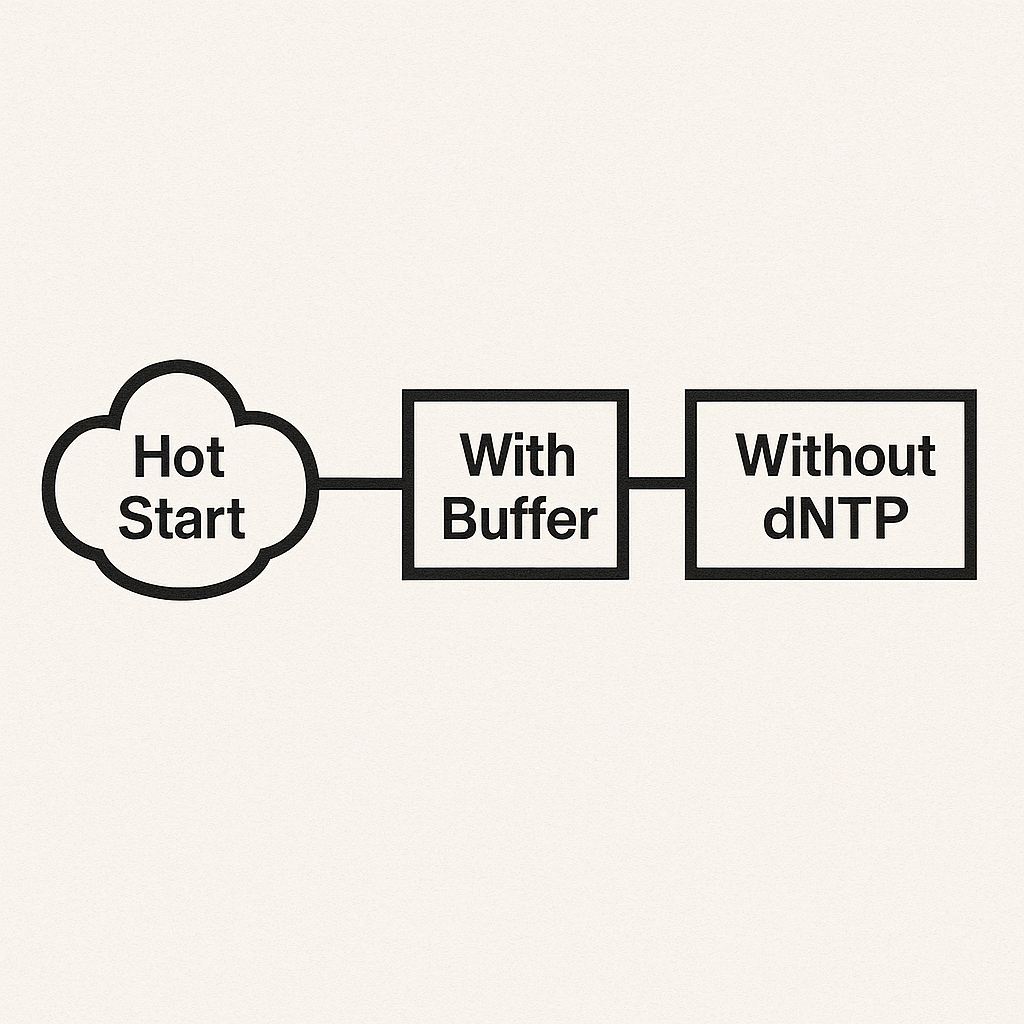
Hot Start DNA Polymerase (With Buffer, With MgCl₂, Without dNTP): Technical Overview, Molecular Mechanisms, Performance Characteristics, and Research Applications

Introduction

Hot Start DNA Polymerase (with Buffer, with MgCl₂, without dNTP) is a specialized PCR formulation designed to improve amplification specificity, reduce nonspecific background, and enable robust amplification from complex templates. This reagent configuration is widely used in qPCR, endpoint PCR, high-fidelity amplification, genotyping, sequencing library preparation, and low-copy detection assays.

The formulation consists of:

  • A Hot Start enzyme (chemically or antibody-inactivated)

  • A ready-to-use reaction buffer

  • MgCl₂ supplied at an optimized concentration (commonly 1.5–3.0 mM)

  • No dNTPs included, giving the user flexibility over nucleotide concentration and purity

This article provides a complete technical overview of molecular mechanisms, assay optimization, reaction chemistry, enzyme activation, and the practical implications of providing buffer + MgCl₂ but excluding dNTPs.

AffiTaq® Hot Start - With Buffer - With MgCl2 - Without dNTP

What Is Hot Start DNA Polymerase?

Hot Start polymerases are thermally activated DNA polymerases engineered to remain inactive at room temperature. This prevents:

  • Nonspecific primer binding

  • Primer-dimer formation

  • Low-temperature polymerase activity

  • Background amplification before denaturation

Hot Start systems use:

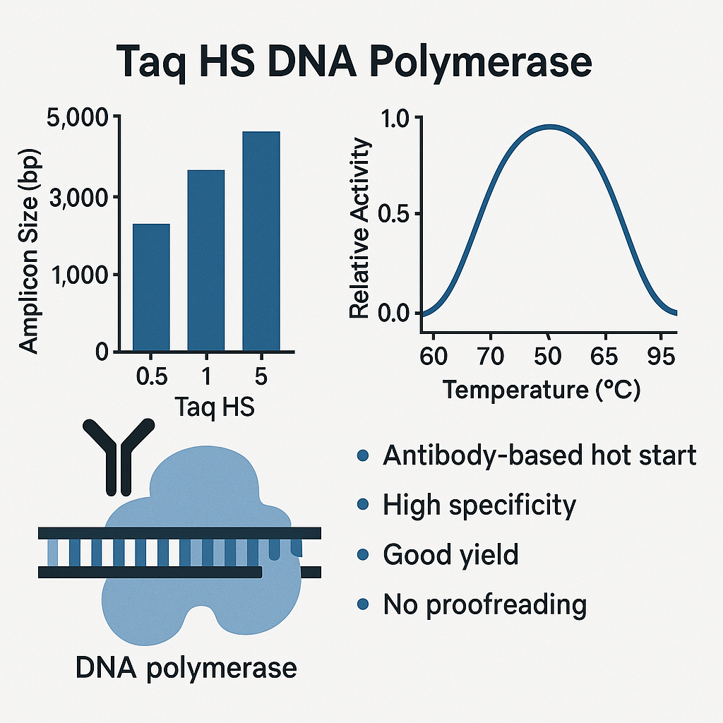
1. Antibody-mediated inhibition

An antibody blocks polymerase activity until heat denaturation (approx. 95 °C).

2. Chemical modification

Polymerase is chemically blocked and reactivated by heat.

3. Aptamer-based inhibition

A nucleic acid aptamer binds the active site until high-temperature activation.

4. Proprietary reversible inhibitors

Often used in commercial PCR master mixes.

The enzyme becomes active only after initial denaturation, improving specificity and sensitivity.

Why Include Buffer and MgCl₂, but Exclude dNTPs?

This configuration is preferred in advanced PCR workflows.

Advantages of Buffer + MgCl₂ Included

  • Ensures reproducibility between reactions

  • Maintains optimized ionic strength

  • Supports consistent pH and enzyme activity

  • Eliminates pipetting variability

Advantages of Excluding dNTPs

Allows full experimental control:

  • User can adjust dNTP concentration for low-copy templates

  • Enables incorporation of specialized dNTP analogs (e.g., dUTP, fluorescent dNTPs)

  • Supports qPCR, mutagenesis, or NGS library preparation protocols requiring custom dNTP ratios

  • Enables compatibility with UNG digestion systems (dUTP incorporation)

Not including dNTPs increases flexibility for:

  • Multiplex PCR

  • Long-range PCR

  • GC-rich or AT-rich genomes

  • High-fidelity proofreading enzymes

Reaction Buffer Composition

A standard hot start buffer typically contains:

  • Tris-HCl (pH 8.3–8.8)

  • KCl or ammonium sulfate

  • MgCl₂ (bound or free)

  • Stabilizers for thermal protection

  • Non-ionic detergents (Tween-20, NP-40)

  • Reducing agents (DTT)

  • Enzyme stabilizing proteins

The buffer ensures enzyme stability at denaturation temperatures and consistency across replicates.

Role of MgCl₂ in Hot Start PCR

Mg²⁺ is an essential cofactor for DNA polymerases.

Functions:

  • Stabilizes dNTP binding

  • Coordinates the active site geometry

  • Enhances polymerase processivity

  • Influences Tm and primer annealing behavior

Effects of Mg²⁺ concentration:

MgCl₂ (mM) Effect
Low (<1.5 mM) Weak yield, low efficiency
Optimal (1.5–3.0 mM) High yield, specific amplification
High (>4.0 mM) Non-specific amplification, mispriming

Providing MgCl₂ pre-optimized ensures consistent reaction performance.

Why dNTPs Are Not Included

This design provides maximum flexibility for:

1. High-fidelity or proofreading polymerases

Some require modified dNTP concentrations.

2. qPCR workflows with fluorescent probes

Probe degradation can occur with excessive dNTP concentrations.

3. NGS and mutagenesis workflows

Precise dNTP stoichiometry is crucial.

4. Use of modified nucleotides

Examples:

  • dUTP

  • Cy5-dCTP

  • Locked nucleic acid (LNA) mixes

  • 7-deaza-GTP for GC-rich templates

5. UV-crosslinking contamination control systems

UNg (uracil-N-glycosylase) requires dUTP incorporation, which must be user-defined.

Mechanism of Activation (Hot Start Release)

Activation occurs during the initial denaturation step, typically:

  • 95 °C

  • 2–10 minutes (depending on inhibitor type)

Activation steps:

  1. Temperature elevation disrupts inhibitor-polymerase interaction

  2. Polymerase undergoes conformational shift

  3. Active catalytic site becomes accessible

  4. Amplification begins during annealing/extension cycles

This prevents premature polymerase activity, increasing amplification fidelity.

Performance Characteristics

1. High Specificity

No polymerase activity at ≤25 °C.

2. High Sensitivity

Detects low-copy targets with minimal background.

3. Consistent Amplification

Buffer and MgCl₂ ensure run-to-run consistency.

4. Improved Reproducibility

Ideal for clinical workflows, forensic DNA, and environmental low-biomass samples.

5. Wide Template Compatibility

Works with:

  • Genomic DNA

  • Plasmids

  • Viral DNA

  • Bacterial DNA

  • cDNA

Applications

Clinical Research

  • Detection of pathogen DNA

  • High-specificity diagnostics

  • SNP genotyping

  • Copy-number variation (CNV) analysis

Molecular Biology

  • Cloning, plasmid verification

  • GC-rich template PCR

  • Mutagenesis

  • Sequencing library prep

qPCR / Real-Time PCR

  • SYBR Green assays

  • TaqMan assays

  • Probe-based quantification

Environmental & Food Testing

  • Pathogen screening

  • Water microbiology

  • GMO detection

Forensics

  • Low-template amplification

  • Touch DNA workflows

Troubleshooting When Using Hot Start Polymerase (Buffer + MgCl₂, No dNTP)

Weak or No Amplification

  • Insufficient MgCl₂ (increase by 0.5 mM increments)

  • Low dNTP concentration

  • Incorrect buffer ratio

  • Poor primer design

Nonspecific Bands

  • MgCl₂ too high

  • Annealing temperature too low

  • Excessive primer concentration

High Ct in qPCR

  • Degradation of dNTPs

  • Impure template

  • Incorrect Mg²⁺:dNTP ratio

Smearing

  • Excessive MgCl₂

  • Template impurities (proteins, salts, EDTA)

Storage and Stability

  • Store at –20 °C

  • Avoid freeze-thaw cycles

  • Protect buffer and MgCl₂ from CO₂ contamination (pH drift)

  • Keep dNTPs separately at –20 °C to prevent hydrolysis

Stability typically ranges from 12–24 months.

Conclusion

Hot Start – With Buffer – With MgCl₂ – Without dNTP is a flexible, high-precision PCR reagent system designed for optimized performance in advanced molecular biology and clinical research. The inclusion of buffer and MgCl₂ ensures reproducibility, while the exclusion of dNTPs allows maximum customization for specialized workflows such as qPCR, high-fidelity PCR, NGS library preparation, and modified nucleotide incorporation.

This configuration supports high specificity, low background, strong amplification efficiency, and compatibility with a broad range of templates and reaction formats.