
Introduction to Paxillin and Quantitative Protein Measurement
The dynamic regulation of cell adhesion, cytoskeletal organization, and intracellular signaling remains central to modern molecular and cellular biology. Among the key scaffold proteins orchestrating these processes is paxillin, a focal adhesion-associated adaptor protein that integrates signals from integrins, growth factor receptors, and cytoskeletal components.
The Paxillin ELISA is a high-sensitivity immunoassay designed for quantitative measurement of paxillin protein levels in biological samples such as cell lysates, tissue extracts, and experimental culture systems. As research increasingly focuses on mechanotransduction, adhesion signaling pathways, and cytoskeletal remodeling, reliable quantification of paxillin has become essential for reproducible data generation.
Educational overviews of focal adhesion biology are available through the National Institutes of Health (NIH) at https://www.nih.gov/ and through cell signaling resources at the National Center for Biotechnology Information (NCBI) https://www.ncbi.nlm.nih.gov/. Structural insights into adhesion proteins are further described by the RCSB Protein Data Bank (PDB) https://www.rcsb.org/, hosted in collaboration with research institutions including Rutgers University.
Understanding Paxillin: Structure and Biological Role
Paxillin is a cytoplasmic phosphoprotein localized at focal adhesions—specialized multi-protein complexes that anchor cells to the extracellular matrix. It contains LD motifs and LIM domains that facilitate interactions with:
-
Focal adhesion kinase (FAK)
-
Src-family kinases
-
Integrins
-
Vinculin
-
Actin-associated proteins
Comprehensive educational materials on cytoskeletal organization can be found at:
-
University of Washington Cell Biology: https://www.biology.washington.edu/
-
Harvard Medical School Cell Biology Program: https://hms.harvard.edu/
-
Stanford School of Medicine Molecular Biology: https://med.stanford.edu/
-
Yale Molecular Biophysics & Biochemistry: https://mbb.yale.edu/
-
University of Michigan Biological Sciences: https://lsa.umich.edu/biology
These academic resources highlight how adaptor proteins like paxillin coordinate signal transduction pathways involved in cellular architecture and environmental sensing.
What Is Paxillin ELISA?
A Paxillin ELISA kit is a quantitative sandwich enzyme-linked immunosorbent assay developed to measure paxillin protein concentration with high specificity and reproducibility.
ELISA (Enzyme-Linked Immunosorbent Assay) remains one of the most widely adopted immunoassay formats in molecular biology laboratories. Methodological foundations are described by:
-
University of California, San Diego (UCSD) Biology Department: https://biology.ucsd.edu/
-
University of Colorado Boulder Biochemistry: https://www.colorado.edu/biochemistry/
-
Cold Spring Harbor Laboratory: https://www.cshl.edu/
-
National Institute of Standards and Technology (NIST): https://www.nist.gov/
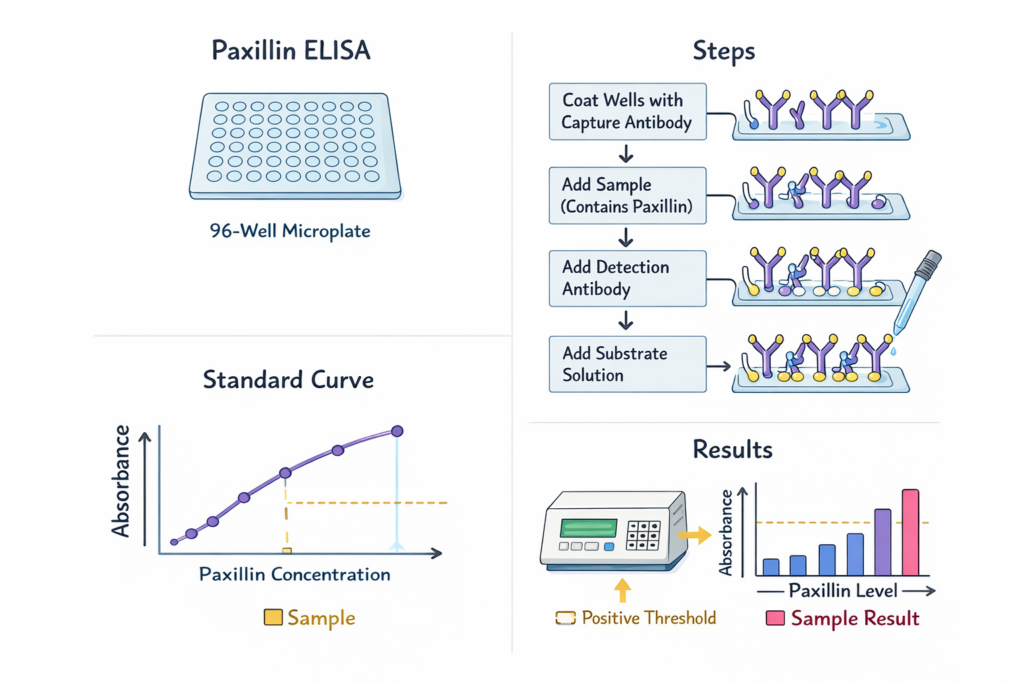
Principle of the Paxillin Sandwich ELISA
The Paxillin ELISA follows a standardized workflow:
1. Capture Phase
Microplate wells are pre-coated with an anti-paxillin capture antibody.
2. Sample Binding
Biological samples are added, allowing paxillin protein to bind specifically.
3. Detection Phase
A secondary enzyme-conjugated detection antibody recognizes a separate epitope.
4. Signal Generation
Substrate conversion produces a measurable colorimetric signal proportional to paxillin concentration.
This quantitative immunoassay approach is consistent with laboratory training materials from:
-
University of Minnesota Biological Sciences: https://cbs.umn.edu/
-
Texas A&M University Laboratory Protocols: https://vetmed.tamu.edu/
-
National Institute of General Medical Sciences (NIGMS): https://www.nigms.nih.gov/
![AffiELISA® Rat Paxillin ELISA [ Pxn]](https://affigen.com/cdn/shop/files/5BAFG-E4345_5D_20AffiELISA_C2_AE_20Cattle_20IFNg_20Kit_20High-Resolution_20Interferon_20Gamma_20ELISA_20Detection_04a43d3d-0d84-465a-9fd9-c89da52f8274_535x.png?v=1712850933)
Why Quantify Paxillin?
Quantitative analysis of paxillin supports research in:
-
Cell adhesion dynamics
-
Integrin signaling pathways
-
Cytoskeletal remodeling
-
Mechanobiology
-
Experimental oncology models
-
Stem cell differentiation systems
-
Tissue engineering research
The NIH Office of Research Infrastructure Programs discusses cytoskeletal signaling research at https://orip.nih.gov/, while mechanobiology research initiatives are described by the National Science Foundation (NSF) at https://www.nsf.gov/.
Paxillin in Focal Adhesion Research
Focal adhesions serve as communication hubs between the extracellular matrix and intracellular signaling cascades. Paxillin functions as a scaffold that recruits regulatory kinases and structural proteins.
Educational discussions of adhesion complexes are available at:
-
University of Cambridge Department of Biochemistry: https://www.bioc.cam.ac.uk/
-
Johns Hopkins Cell Biology: https://cellbio.jhmi.edu/
-
National Library of Medicine (NLM): https://www.nlm.nih.gov/
-
National Human Genome Research Institute (NHGRI): https://www.genome.gov/
Advantages of Paxillin ELISA Over Alternative Techniques
Compared with other protein detection techniques such as Western blotting or mass spectrometry, Paxillin ELISA provides:
-
High-throughput quantification
-
Reduced assay variability
-
Objective numerical output
-
Scalability for multi-sample studies
-
Streamlined protocol workflow
Standardization practices are supported by the CDC Laboratory Quality Management Systems https://www.cdc.gov/labquality/ and research reproducibility guidelines from the NIH Office of Extramural Research https://grants.nih.gov/.
Sample Types Compatible with Paxillin ELISA
The Paxillin ELISA kit is suitable for:
-
Cultured cell lysates
-
Tissue homogenates
-
Experimental protein extracts
-
Conditioned media (depending on protocol)
Best practices in sample preparation are described in academic resources from:
-
University of Wisconsin Biotechnology Center: https://biotech.wisc.edu/
-
University of North Carolina Lineberger Research Resources: https://unclineberger.org/
-
National Cancer Institute Research Methods Portal: https://www.cancer.gov/research
Data Analysis and Quantitative Interpretation
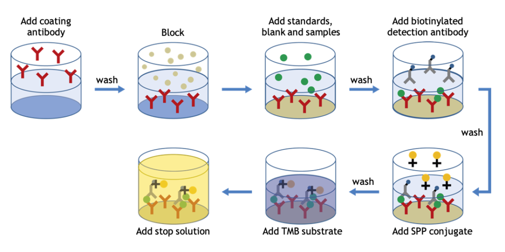
ELISA data are analyzed using a standard curve derived from known concentrations of paxillin standards. Concentrations are typically expressed in ng/mL or pg/mL.
Statistical guidance for assay validation and quantitative reproducibility can be found at:
-
National Institute of Statistical Sciences: https://www.niss.org/
-
NIH Biostatistics Resources: https://www.nhlbi.nih.gov/
-
U.S. Department of Energy Office of Science Biological Research: https://science.osti.gov/
Research Areas Supported by Paxillin ELISA
1. Cell Migration Studies
2. Integrin Signaling Analysis
3. Cytoskeletal Remodeling Research
4. Tumor Microenvironment Models
5. Tissue Engineering and Biomaterials Research
The National Institute of Biomedical Imaging and Bioengineering (NIBIB) provides related educational resources at https://www.nibib.nih.gov/.
-
Paxillin ELISA kit
-
quantitative paxillin assay
-
focal adhesion protein analysis
-
cell signaling ELISA
-
integrin pathway quantification
-
scaffold protein measurement
-
high sensitivity sandwich ELISA
-
laboratory immunoassay kit
-
protein expression profiling assay
These keywords align with commonly searched terms in molecular biology and cell signaling research.
Best Laboratory Practices
To ensure reliable results:
✔ Run duplicates or triplicates
✔ Include internal controls
✔ Maintain consistent incubation timing
✔ Use calibrated pipettes
✔ Validate reagent storage conditions
Laboratory quality standards are discussed by:
-
National Institutes of Health Research Training: https://researchtraining.nih.gov/
-
Occupational Safety and Health Administration (OSHA) Laboratory Safety: https://www.osha.gov/laboratory-safety
-
NIH Biosafety Guidelines: https://osp.od.nih.gov/biotechnology/
Expanding Applications in Mechanobiology and Systems Biology
As integrative biology expands, quantitative assays like Paxillin ELISA support systems-level analysis of signaling networks. Educational initiatives from:
-
National Science Foundation BIO Directorate: https://www.nsf.gov/bio/
-
NIH Systems Biology Programs: https://commonfund.nih.gov/
highlight the importance of scaffold proteins in complex regulatory networks.
Conclusion
The Paxillin ELISA represents a robust, quantitative tool for investigating focal adhesion dynamics and intracellular signaling pathways. By enabling accurate measurement of paxillin protein levels, researchers can better understand cytoskeletal organization, integrin-mediated signaling, and cellular architecture regulation.
Supported by standardized methodology and referenced by leading academic and government institutions, Paxillin ELISA remains a valuable component of modern cell biology research workflows.