
Introduction to TBX18 and Antibody-Based Analysis
Understanding how transcription factors regulate gene expression is fundamental to cell biology, developmental biology, and functional genomics. TBX18 (T-box transcription factor 18) is one such regulatory protein, involved in embryonic development and tissue differentiation across multiple systems.
To support studies of TBX18 expression and function, the Anti-TBX18 Polyclonal IgG Antibody is widely used as a research reagent to detect TBX18 in applications such as Western blotting, immunocytochemistry (ICC), immunohistochemistry (IHC), and enzyme-linked immunosorbent assays (ELISA).
Foundational background on transcription factors can be explored at the National Center for Biotechnology Information (NCBI): https://www.ncbi.nlm.nih.gov/
General transcription factor roles are described by the National Institutes of Health (NIH): https://www.nih.gov/
Additional insights into developmental biology and gene regulation are provided by Harvard University’s Department of Molecular and Cellular Biology: https://mcb.harvard.edu/
What Is TBX18?
TBX18 belongs to the T-box family of transcription factors — a conserved group of DNA-binding proteins that orchestrate gene expression during embryogenesis and cellular differentiation.
Key characteristics of TBX18:
-
DNA-binding transcription factor
-
Expressed in specific embryonic tissues
-
Involved in organogenesis
-
Regulates gene networks linked to development
T-box proteins and their roles are further explained by developmental biology resources, such as the Society for Developmental Biology (SDB): https://www.sdbonline.org/
Genetic and protein data related to TBX18 are accessible via the European Bioinformatics Institute (EMBL-EBI): https://www.ebi.ac.uk/
What Is a Polyclonal IgG Antibody?
An antibody is an immune-derived protein used by researchers to specifically recognize and bind a target antigen — in this case, TBX18.
Polyclonal IgG antibodies are a mixture of immunoglobulin G molecules that bind multiple epitopes on the same antigen. Benefits of polyclonal IgG include:
-
Recognition of multiple protein regions
-
Enhanced detection sensitivity
-
Robust performance across various sample types
The structure and function of IgG antibodies are described by immunology training resources such as the American Association of Immunologists (AAI): https://www.aai.org/
Principles of antibody specificity and binding are detailed in educational material from the University of California, San Diego (UCSD): https://biology.ucsd.edu/
Why Use Anti-TBX18 Polyclonal IgG in Research?
1. Protein Expression Profiling
Anti-TBX18 antibodies can be used to detect endogenous TBX18 protein levels in cells and tissues, providing insight into regulatory pathways and transcriptional networks.
Understanding protein regulation is a core topic in molecular biology, as described by Stanford University’s Molecular Genetics program: https://med.stanford.edu/
2. Developmental Biology Studies
TBX18 plays a role in early development, making its detection valuable in embryology research.
Developmental gene regulation is covered in Yale University’s developmental biology resources: https://medicine.yale.edu/
3. Cellular Localization (ICC, IHC)
Antibody-based imaging techniques allow researchers to visualize where TBX18 is localized within cells or tissue architecture.
Guidance on immunocytochemistry and immunohistochemistry techniques is available from Cold Spring Harbor Laboratory (CSHL): https://www.cshl.edu/
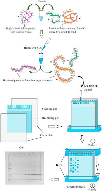
4. Electrophoretic Protein Analysis
Anti-TBX18 antibodies are suitable for Western blot applications, enabling quantification of TBX18 levels under different experimental conditions.
Protein analysis principles are taught at the University of Michigan Department of Biochemistry: https://lsa.umich.edu/biochemistry/
Applications and Assay Formats
Western Blotting (WB)
Used to separate and detect TBX18 by molecular weight after gel electrophoresis.
Learn more about protein blotting techniques from the National Institute of General Medical Sciences (NIGMS): https://www.nigms.nih.gov/
Immunocytochemistry (ICC) & Immunofluorescence
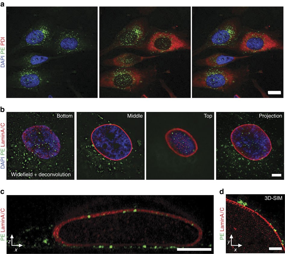
Enables visualization of TBX18 within fixed cells using antibody labeling and microscopy.
ICC fundamentals are discussed by the University of Iowa Immunology Program: https://medicine.uiowa.edu/igb/
Immunohistochemistry (IHC)
Detects TBX18 expression in fixed tissue sections, useful in developmental biology and histological studies.
IHC methodology is described in detail by the Centers for Disease Control and Prevention (CDC): https://www.cdc.gov/
Enzyme-Linked Immunosorbent Assay (ELISA)
Quantifies TBX18 levels using antibody capture and detection systems.
Foundational immunoassay reference material is available from the University of Minnesota ELISA training portal: https://cbs.umn.edu/
Choosing the Right Anti-TBX18 Polyclonal IgG Antibody
When selecting an antibody for research use, consider:
✔ Host species (e.g., rabbit, goat) for secondary antibody compatibility
✔ Assay compatibility (WB, ICC, IHC, ELISA)
✔ Validated applications and published citations
✔ Positive and negative controls in experimental design
Antibody validation principles are outlined by the National Institutes of Health Office of Research Quality: https://grants.nih.gov/
Best Practices for Antibody-Based Detection
1. Optimize Antibody Dilution
Test multiple antibody dilutions to determine optimal signal-to-noise ratio.
Immunodetection optimization is covered in training resources from Texas A&M University: https://vetmed.tamu.edu/
2. Proper Controls
Include positive controls that express TBX18 and negative controls lacking the target protein.
Learn more about control strategies from the University of Wisconsin Biotechnology Center: https://biotech.wisc.edu/
3. Blocking and Washing
Effective blocking and rigorous washes reduce background signal and improve specificity.
Blocking strategies and wash methods are described by the University of California, Davis (UC Davis) Immunology Program: https://www.ucdavis.edu/
4. Secondary Antibody Selection
Match the secondary antibody species to the host of the polyclonal IgG for enhanced detection.
Secondary antibody fundamentals are detailed by Harvard Medical School’s Immunology Program: https://immunology.hms.harvard.edu/
Interpretation of Results
Positive detection with Anti-TBX18 Polyclonal IgG antibody indicates the presence and relative abundance of TBX18 protein under experimental conditions. Interpretation should be aligned with:
-
Experiment design and controls
-
Exposure or signal intensity
-
Comparative sample sets
Quantitative analysis approaches are further outlined by the National Institute of Statistical Sciences (NISS): https://www.niss.org/
Research Tools and Resources
To support antibody-based research, the following educational resources provide foundational knowledge:
-
NIH Gene Database (NCBI) – https://www.ncbi.nlm.nih.gov/gene
-
Protein Data Bank (PDB) – https://www.rcsb.org/
-
Gene Expression Resources (EMBL-EBI) – https://www.ebi.ac.uk/
-
Immunology Learning Modules (AAI) – https://www.aai.org/
-
Cell Biology Resources (Harvard MCB) – https://mcb.harvard.edu/
SEO-Driven Keywords Integrated Naturally
-
Anti-TBX18 Polyclonal IgG antibody
-
TBX18 protein detection
-
transcription factor research reagent
-
Western blot antibody TBX18
-
immunocytochemistry TBX18
-
immunohistochemistry antibody research
-
ELISA detection of TBX18
-
polyclonal IgG research antibody
-
gene regulatory protein assay
Conclusion
The Anti-TBX18 Polyclonal IgG Antibody is a versatile and essential research reagent for studying TBX18 transcription factor expression, localization, and regulation in cellular and developmental biology. Its broad compatibility with key research methods — including Western blot, ICC, IHC, and ELISA — makes it valuable for scientists investigating gene regulation, differentiation pathways, and protein expression networks.
By combining robust antibody validation practices, appropriate controls, and rigorous experimental design, researchers can achieve clear, reproducible insights into TBX18-associated biology.