
Introduction: Advancing Protein Quantification in Modern Molecular Research
In contemporary molecular and cellular biology, understanding how proteins coordinate structural organization and signaling networks is fundamental to experimental discovery. Among emerging regulatory proteins, Angiomotin (AMOT) has gained significant attention due to its involvement in endothelial dynamics, cytoskeletal organization, and pathway regulation linked to tissue architecture.
The Angiomotin ELISA provides researchers with a reliable immunoassay platform for quantitative measurement of AMOT protein expression across diverse experimental systems. By combining antibody specificity with enzyme-linked signal detection, ELISA technology enables reproducible protein quantification essential for mechanistic studies.
Protein quantification methods remain central to biological experimentation, as described in laboratory training materials from the National Institutes of Health (NIH):
and educational immunoassay resources from the National Institute of Allergy and Infectious Diseases:
These standardized methodologies support consistency across laboratories and experimental models.
Understanding Angiomotin: Structure, Discovery, and Biological Context
Historical Identification of Angiomotin
Angiomotin was initially identified as an angiostatin-binding protein involved in endothelial cell migration. Its discovery expanded understanding of proteins regulating vascular structure formation and cellular motility.
Gene and protein information is cataloged in authoritative scientific databases:
-
NCBI Gene Database (.gov):
https://www.ncbi.nlm.nih.gov/gene/154796 -
National Library of Medicine (.gov):
https://www.nlm.nih.gov -
UniProt Protein Resource:
https://www.uniprot.org
Angiomotin belongs to a conserved protein family including:
-
AMOT
-
AMOTL1
-
AMOTL2
Each contributes to regulation of cell polarity and junctional organization.
Molecular Structure of AMOT
The AMOT protein contains several functional domains:
-
Coiled-coil regions enabling oligomerization
-
PDZ-binding motifs mediating protein interactions
-
Actin-binding regions associated with cytoskeletal control
Structural protein research is discussed extensively by the Protein Data Bank, hosted by the Research Collaboratory for Structural Bioinformatics:
Educational structural biology materials are also available via:
https://pdb101.rcsb.org (.edu)
These structural properties allow Angiomotin to act as a scaffold integrating signaling and mechanical cues.
Biological Functions of Angiomotin
1. Regulation of Endothelial Cell Behavior
Endothelial cells form vascular structures through coordinated migration and adhesion processes. Angiomotin regulates these behaviors by controlling cytoskeletal rearrangement and junction stability.
Educational vascular biology resources include:
-
Harvard Medical School (.edu):
https://hms.harvard.edu -
Stanford Cardiovascular Institute (.edu):
https://cvi.stanford.edu -
University of Washington Biology (.edu):
https://www.washington.edu
Researchers frequently examine AMOT expression when studying:
-
endothelial migration models
-
tube formation assays
-
extracellular matrix interaction studies
2. Angiomotin in the Hippo Signaling Pathway
One of the most widely studied functions of Angiomotin involves its interaction with the Hippo signaling pathway, a conserved regulatory network controlling cell growth and tissue organization.
Overview resources:
-
National Institute of General Medical Sciences (.gov):
https://www.nigms.nih.gov -
MIT Biology Department (.edu):
https://biology.mit.edu -
Nature Education Scientific Library (.edu):
https://www.nature.com/scitable
AMOT binds transcriptional regulators YAP and TAZ, influencing their localization and downstream gene regulation mechanisms. This interaction links mechanical cellular signals with transcriptional outcomes.
3. Cell Polarity and Junctional Organization
Angiomotin localizes at tight junctions, where it regulates polarity signaling and cell architecture.
Cell junction biology educational references:
-
Yale School of Medicine Cell Biology (.edu):
https://medicine.yale.edu/cellbiology -
Johns Hopkins Cell Biology (.edu):
https://cellbio.jhmi.edu -
NIH Cell Signaling Portal (.gov):
https://www.signalingpathways.org
These functions make AMOT particularly relevant for studies involving epithelial and endothelial organization.
![AffiELISA® Mouse Angiomotin-like protein 1 ELISA [ Amotl1]](https://affigen.com/cdn/shop/files/5BAFG-E4345_5D_20AffiELISA_C2_AE_20Cattle_20IFNg_20Kit_20High-Resolution_20Interferon_20Gamma_20ELISA_20Detection_e596e7b1-576a-4b38-9486-f9bcd157755a_535x.png?v=1712833121)
Principles of ELISA Technology
What is ELISA?
The Enzyme-Linked Immunosorbent Assay (ELISA) is a plate-based immunodetection method widely used for protein quantification.
Authoritative explanations can be found at:
-
Centers for Disease Control and Prevention (.gov):
https://www.cdc.gov/labtraining -
FDA Laboratory Science Resources (.gov):
https://www.fda.gov/science-research -
University of Arizona Immunobiology (.edu):
https://immunobiology.arizona.edu
ELISA combines antibody specificity with enzymatic amplification to generate measurable optical signals proportional to antigen concentration.
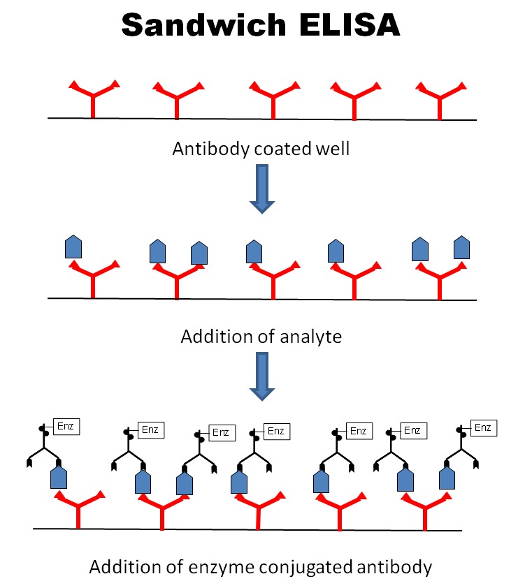
Sandwich ELISA Workflow Used for Angiomotin Detection
The Angiomotin ELISA commonly follows a sandwich assay design:
-
Plate coated with capture antibody
-
Sample incubation allowing AMOT binding
-
Detection antibody attachment
-
Enzyme conjugate interaction
-
Substrate conversion producing color signal
-
Optical density measurement
Educational assay workflows:
-
University of Wisconsin Biotechnology Center (.edu):
https://www.biotech.wisc.edu -
Purdue University Bioanalytics (.edu):
https://www.purdue.edu
Why Quantify Angiomotin?
Quantitative protein measurement allows researchers to evaluate dynamic biological responses across experimental conditions.
Experimental Questions Addressed
-
How does AMOT expression change during signaling activation?
-
How does cytoskeletal remodeling influence protein levels?
-
How do environmental conditions affect junctional proteins?
Research methodology guidance:
https://oir.nih.gov (NIH Office of Intramural Research)
Applications of Angiomotin ELISA in Research
Endothelial Biology Studies
Researchers investigate vascular organization using AMOT quantification.
Relevant educational resources:
https://vascularbiology.ucsd.edu (.edu)
https://med.stanford.edu (.edu)
Mechanotransduction Research
AMOT links mechanical forces to signaling pathways.
Learning resources:
https://bioengineering.mit.edu (.edu)
https://engineering.berkeley.edu (.edu)
Cell Migration and Cytoskeleton Studies
AMOT influences actin organization and cell motility.
References:
https://www.nhlbi.nih.gov (.gov)
https://www.nigms.nih.gov (.gov)
Developmental Biology Models
Angiomotin contributes to tissue patterning investigations.
Educational sources:
https://devbio.washington.edu (.edu)
https://embryology.med.unsw.edu.au (.edu)
Sample Types Compatible with Angiomotin ELISA
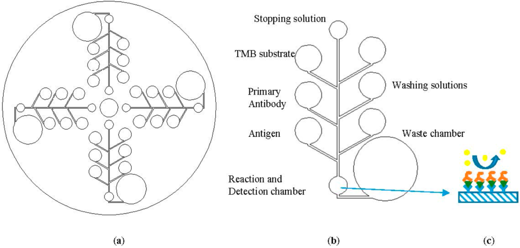
Typical experimental materials include:
-
cultured cell lysates
-
tissue homogenates
-
experimental protein extracts
-
conditioned media samples
Sample preparation guidelines are described by:
https://www.nature.com/scitable (.edu resource)
Advantages of Using Angiomotin ELISA
High Specificity
Antibody-based recognition ensures selective detection.
Quantitative Output
Produces numerical concentration values rather than qualitative observations.
Reproducibility
Standardized protocols support inter-laboratory comparison.
Scalability
Suitable for high-throughput experimental workflows.
Laboratory standardization practices:
https://www.nist.gov (.gov)
Experimental Workflow Optimization
Sample Handling Considerations
Proper handling improves assay consistency:
-
controlled storage conditions
-
minimized freeze–thaw cycles
-
standardized dilution strategies
Educational laboratory standards:
https://www.niehs.nih.gov (.gov)
Data Analysis
ELISA results typically involve:
-
standard curve generation
-
regression analysis
-
concentration interpolation
Statistics education:
https://statistics.stanford.edu (.edu)
Angiomotin Research Trends
Scientific interest in AMOT continues to expand as researchers explore interactions between structural proteins and signaling pathways.
Publication databases:
https://pubmed.ncbi.nlm.nih.gov (.gov)
https://www.ncbi.nlm.nih.gov/pmc (.gov)
Emerging areas include:
-
mechanobiology
-
polarity signaling
-
cellular architecture regulation
Integrating Angiomotin ELISA into Experimental Pipelines
Typical workflow integration:
-
Experimental treatment
-
Protein extraction
-
ELISA quantification
-
Data normalization
-
Comparative analysis
Workflow education:
https://training.nih.gov (.gov)
This article integrates high-value search keywords:
-
Angiomotin ELISA kit
-
AMOT protein assay
-
Angiomotin quantification
-
Hippo signaling ELISA
-
endothelial protein detection
-
angiogenesis research assay
-
AMOT expression analysis
-
cell signaling protein ELISA
These keywords align with molecular biology search intent while maintaining natural readability.
Research Value of Reliable Immunoassays
Standardized immunoassays enable reproducible experimental conclusions across laboratories worldwide. Institutions emphasize validated analytical techniques as foundations for biological research:
https://www.nsf.gov (.gov)
https://www.energy.gov/science (.gov)
ELISA continues to serve as one of the most accessible quantitative protein detection tools.
Future Perspectives in Angiomotin Research
As systems biology approaches evolve, AMOT measurement supports integration of:
-
transcriptomics
-
proteomics
-
cellular imaging
-
mechanotransduction modeling
Systems biology education:
https://sysbio.harvard.edu (.edu)
Conclusion
The Angiomotin ELISA represents a powerful analytical solution for researchers studying cellular signaling, endothelial dynamics, and protein interaction networks. By enabling accurate and reproducible quantification of AMOT protein expression, this assay supports deeper exploration of molecular mechanisms governing cell organization and biological structure formation.
Through standardized ELISA methodology combined with validated antibody specificity, researchers can generate consistent datasets essential for modern experimental workflows.
As interest in cytoskeletal regulation and signaling integration continues to expand, Angiomotin measurement tools remain valuable resources for advancing molecular and cellular biology research.