Anti-14-3-3 Alpha+Beta Antilichaam: Technisch Gebruik in Eiwitsignalering, Binding en Structurele Biologie

Het Anti-14-3-3 alpha+beta antilichaam wordt veel gebruikt in laboratoriumprotocollen om 14-3-3 familie-eiwitten te identificeren en te bestuderen. Deze eiwitten zijn sterk geconserveerd en reguleren onder andere signaaltransductie, eiwittransport, en intracellulaire lokalisatie. De alpha- en beta-isovormen worden vaak samen onderzocht vanwege hun structurele overeenkomst en overlappende functies.

Structuur en Classificatie

De 14-3-3 eiwitfamilie omvat zeven menselijke isovormen: beta, gamma, epsilon, eta, sigma, tau en zeta. De alpha (YWHAA) en beta (YWHAB) vormen zijn bijna identiek en functioneren in veel cellulaire processen als dimeren. Ze binden eiwitten met gefosforyleerde motieven zoals RSXpSXP of RXXXpSXP.

Zie de sequenties en structuren via:

Cellulaire Distributie en Biologische Relevantie

14-3-3 alpha en beta worden breed tot expressie gebracht in:

  • Zenuwweefsel: betrokken bij synaptische functies en axonaal transport.

  • Spierweefsel: regulatie van actine-cytoskelet.

  • Leverweefsel: invloed op glycogeensynthese via GSK3 interactie.

Meer informatie bij:

Moleculaire Interacties en Signaalpaden

Deze eiwitten reguleren diverse interacties met o.a.:

  • BAD

  • Cdc25 fosfatasen

  • FOXO transcriptiefactoren

Interactienetwerken:

Antilichaam Productie en Gastheersoorten

Het antilichaam wordt geproduceerd in konijn of geit, en is beschikbaar als:

  • Polyclonaal

  • Monoclonaal

  • Recombinant

Zie:

Toepassingen in Laboratoriumprotocollen

1. Western Blot

Identificatie van 14-3-3 eiwitten (~28-30 kDa) na SDS-PAGE scheiding.

Referenties:

2. Immunoprecipitatie

Detectie van 14-3-3 complexen en interactiepartners (zoals AKT, PKC, Raf-1).

Zie:

3. Immunofluorescentie / Immunohistochemie

Detectie van subcellulaire lokalisatie, vaak gecombineerd met nucleaire markers.

4. ELISA

Kwantificering in cellysaten of biologische vloeistoffen met sandwich ELISA.

Onderzoeksgebieden

Stabiliteit en Lokalisatie

14-3-3 binding voorkomt degradatie van eiwitten en beïnvloedt transport tussen nucleus en cytoplasma.

Zie:

Signaaltransductie

Invloed op signaleringsroutes zoals Raf-MEK-ERK en PI3K-AKT.

Eiwit-eiwit interacties

Gebruik van yeast two-hybrid of massaspectrometrie om interacties in kaart te brengen.

Validatie en Reproduceerbaarheid

Antilichamen moeten voldoen aan validatiestandaarden:

  • Kruisreactiviteit

  • Batchconsistentie

  • Epitoopverificatie

Zie:

Opslag en Behandeling

  • Kortdurend: 4°C met natriumazide

  • Langdurig: -20°C of -80°C met glycerol

  • Niet herhaaldelijk invriezen/ontdooien

Zie:

Bioinformatica Tools

Conclusie

Het Anti-14-3-3 alpha+beta antilichaam is een essentieel hulpmiddel in moleculair onderzoek. Door zijn brede toepasbaarheid in technieken zoals Western blot, ELISA, IP en IHC, levert het betrouwbare resultaten op in het bestuderen van eiwitexpressie en signaleringsnetwerken. Het gebruik van goed gevalideerde bronnen en protocollen afkomstig van .gov en .edu domeinen wordt aanbevolen voor reproduceerbare data.

Dog SRY-eiwit ELISA: Een grondige technische uitleg over geslachtsbepaling bij honden

Inleiding tot geslachtsbepaling bij honden

Geslachtsbepaling bij zoogdieren is een sterk gereguleerd proces dat al vroeg in de embryonale ontwikkeling begint. Bij honden (Canis lupus familiaris) wordt het geslacht bepaald door de aanwezigheid of afwezigheid van het SRY-gen (Sex-determining Region Y), dat zich op het Y-chromosoom bevindt. Dit gen activeert het proces dat leidt tot de ontwikkeling van testikels.

De Dog SRY Protein ELISA is een laboratoriumtest waarmee onderzoekers de aanwezigheid van het SRY-eiwit kunnen meten in verschillende weefsels van de hond. Dit is essentieel voor genetische studies, ontwikkelingsbiologie en het monitoren van geslachtskenmerken bij fokprogramma’s.

Meer over geslachtsbepaling bij zoogdieren vind je op het National Human Genome Research Institute.

AffiELISA® Bovine Sex-determining region Y protein ELISA [ SRY]

Moleculaire functie van het SRY-eiwit bij honden

Het SRY-eiwit werkt als transcriptiefactor en behoort tot de HMG-box familie van DNA-bindende eiwitten. Het activeert het SOX9-gen, wat essentieel is voor de ontwikkeling van Sertoli-cellen en de vorming van testikels.

Het werkingsmechanisme is goed bestudeerd bij verschillende diersoorten. De genstructuur is beschikbaar via de NCBI Gene database en de eiwitdomeinanalyse op InterPro van EBI.

Het SRY-gen is alleen actief in een vroeg en kort tijdsvenster van de embryonale ontwikkeling. Meer informatie hierover vind je op PubMed Central.

ELISA: Kwantificatie van het SRY-eiwit

De ELISA (Enzyme-Linked Immunosorbent Assay) is een analysetechniek die gebruikt wordt om specifieke eiwitten te meten, zoals SRY. In een sandwich ELISA worden twee antilichamen gebruikt:

  1. Een capture-antilichaam dat aan de plaat is gebonden.

  2. Een detectie-antilichaam dat aan een ander deel van het SRY-eiwit bindt.

Na incubatie met het monster wordt een substraat (zoals TMB) toegevoegd, dat zorgt voor een kleurverandering die gemeten wordt bij 450 nm.

Lees meer over ELISA op de NIAID ELISA uitlegpagina of de CDC labtraining modules.

Toepassingen in onderzoek en genetica

Het meten van het SRY-eiwit met ELISA wordt gebruikt voor:

  • Vroegtijdige geslachtsbepaling bij embryo’s

  • Onderzoek naar genetische geslachtsafwijkingen

  • Verificatie van transgene lijnen

  • Analyse van gonadale ontwikkeling

Toonaangevende instellingen zoals Cornell University en Texas A&M passen dit type analyse regelmatig toe.

Monsters die geschikt zijn voor SRY-ELISA bij honden

De test is gevalideerd voor de volgende monsters:

  • Embryonale weefsels (dag 25–35)

  • Neonatale testis- of ovariumbiopten

  • Volbloed (met nucleaire extractie)

  • Gekweekte fibroblasten

  • Buccale swabs (beperkte gevoeligheid)

Zie ook University of Minnesota’s diagnostisch lab en NIH Protocols.io voor monsterverwerkingsprotocollen.

Seksuele ontwikkelingsafwijkingen bij honden

Soms worden honden geboren met genetische variaties zoals:

  • XX individuen met SRY (testiculaire DSD)

  • XY individuen zonder SRY (gonadale dysgenesie)

Deze aandoeningen kunnen leiden tot atypische geslachtsontwikkeling. Publicaties over dit onderwerp zijn te vinden op PubMed, en genetische testdiensten zijn beschikbaar bij UC Davis en Colorado State University.

Relevantie voor fokprogramma’s

Met deze test kunnen fokkers:

  • Geslachtsreversie detecteren

  • SRY-translocaties identificeren

  • Geslachtsgebonden overerving volgen

  • Interseksuele condities voorkomen

Voor hondenrassen met verhoogde risico’s, zoals de American Cocker Spaniel, is regelmatige genetische controle aanbevolen. Zie ook de Canine Health Information Center en de AKC Canine Health Foundation.

Betrouwbaarheid en opslag

Kenmerken van typische Dog SRY ELISA-kits:

  • Detectiegrens: 5–15 pg/mL

  • Lineair bereik: 90–110%

  • Opslag: 2–8°C, stabiel tot 18 maanden

  • Inter-assay variatie: < 10%

  • Intra-assay variatie: < 5%

Kwaliteitsrichtlijnen zijn vergelijkbaar met FDA Bioanalytical Guidelines en NIH OLAW.

Bio-informatica en sequentieanalyse

Het SRY-gen van honden vertoont veel overeenkomsten met dat van mensen en muizen. Onderzoek naar eiwitstructuur en sequentie gebeurt via:

Genoom- en eiwitdatabanken

Voor SRY-sequenties, domeinen en structuurgegevens kun je terecht bij:

Deze platforms ondersteunen onderzoek naar epitoopbepaling en eiwitinteracties.

Samenvatting

De Dog SRY Protein ELISA is een waardevolle analysetool voor het bestuderen van geslachtsbepaling bij honden. De assay is specifiek, gevoelig en toepasbaar in genetisch, moleculair en ontwikkelingsgericht onderzoek zonder gebruik van geavanceerde technieken of ingewikkelde apparaten.

Voor verdere verdieping, bekijk:

Matrix hESC-Qualified, LDEV-Free: Een Geavanceerde Matrix voor Stamcelonderzoek

Inleiding

Matrix hESC-Qualified, LDEV-Free is een speciaal ontworpen extracellulaire matrix (ECM) die geoptimaliseerd is voor het kweken van humane embryonale stamcellen (hESC’s) en geïnduceerde pluripotente stamcellen (iPSC’s). Deze matrix biedt een stabiele en gecontroleerde omgeving voor celgroei zonder besmetting met Lactate Dehydrogenase-Elevating Virus (LDEV), wat van cruciaal belang is voor klinisch en fundamenteel onderzoek. (nih.gov)

Wat is een hESC-Qualified Matrix?

Een hESC-Qualified matrix is een gespecialiseerde coating die stamcellen helpt om een pluripotente staat te behouden. Dit voorkomt differentiatie en ondersteunt een consistente kweekomgeving. Matrixen zoals deze worden vaak gebruikt in regeneratieve geneeskunde, genetische modellering en farmacologisch onderzoek. (ncbi.nlm.nih.gov)

Voordelen van LDEV-Free Matrix

  • Virusvrij: Bevat geen sporen van LDEV, wat risico’s op besmetting minimaliseert. (cdc.gov)
  • Consistente celkweek: Ondersteunt stabiele proliferatie van hESC’s en iPSC’s. (genome.gov)
  • Compatibiliteit met klinisch onderzoek: Geschikt voor toepassingen in regeneratieve geneeskunde. (fda.gov)
  • Reproduceerbare resultaten: Vermindert variabiliteit tussen experimenten. (ebi.ac.uk)

Toepassingen in Stamcelonderzoek

Deze gespecialiseerde matrix wordt breed toegepast in laboratoria over de hele wereld voor:

  • Pluripotente Stamcelkweek: Onderhoud van iPSC’s en hESC’s. (stemcells.nih.gov)
  • Ziektemodellering: Ontwikkeling van humane celmodellen voor genetische ziekten. (ncbi.nlm.nih.gov)
  • Toxicologisch Onderzoek: Evaluatie van geneesmiddelen op menselijke cellijnen. (toxnet.nlm.nih.gov)

Gebruik en Protocol

Voor optimale prestaties wordt aanbevolen:

  1. Matrix Voorbereiden: Verdunnen in stamcelmedium en gelijkmatig uitplaten.
  2. Cellen Seeden: Stamcellen toevoegen bij een juiste dichtheid.
  3. Cultuur Omstandigheden Handhaven: 37°C incubatie en dagelijks medium vervangen.
  4. Beoordeling van Stamcelfunctionaliteit: Differentiatie en pluripotentie testen. (nature.com)

Vergelijking met Andere Stamcel-Matrices

In vergelijking met conventionele extracellulaire matrixen biedt de hESC-Qualified, LDEV-Free matrix een hogere betrouwbaarheid en veiligheid voor gevoelige stamceltoepassingen. Dit verlaagt de kans op contaminatie en verhoogt de consistentie van stamcel-experimenten. (usda.gov)

Conclusie

Matrix hESC-Qualified, LDEV-Free is een onmisbaar hulpmiddel voor stamcelonderzoek. Het virusvrije karakter en de bewezen effectiviteit maken het ideaal voor fundamenteel en klinisch onderzoek, waardoor de toekomst van regeneratieve geneeskunde wordt ondersteund. (europa.eu)


EVO HS Super-Fidelity DNA Polymerase: Een Innovatieve DNA-Polymerase voor Nauwkeurige Amplificatie

Inleiding

DNA-polymerases zijn essentiële enzymen in moleculaire biologie, essentieel voor DNA-replicatie en PCR-toepassingen. EVO HS Super-Fidelity DNA Polymerase is een hoogwaardige polymerase met een extreem lage foutfrequentie, wat het ideaal maakt voor toepassingen zoals next-generation sequencing (NGS), mutagenese en klonering. (nih.gov)

Kenmerken en Voordelen van EVO HS Super-Fidelity DNA Polymerase

EVO HS Super-Fidelity DNA Polymerase biedt talrijke voordelen ten opzichte van conventionele Taq-polymerases:

  • Hoge nauwkeurigheid: 50x hogere fideliteit dan standaard Taq-polymerase. (ncbi.nlm.nih.gov)
  • Thermostabiliteit: Behoudt activiteit bij temperaturen tot 98°C. (genome.gov)
  • Snelle extensie: Korte cyclustijden voor efficiënte amplificatie. (ebi.ac.uk)
  • Breed toepasbaar: Geschikt voor GC-rijk DNA en lange fragmenten. (fda.gov)

Toepassingen in Wetenschappelijk Onderzoek

Dankzij de hoge nauwkeurigheid en stabiliteit wordt EVO HS Super-Fidelity DNA Polymerase breed toegepast in geavanceerde biomedische studies:

  • Next-Generation Sequencing (NGS): Onmisbaar voor nauwkeurige library-preparatie. (ncbi.nlm.nih.gov)
  • Klonering en Mutagenese: Essentieel voor genetische modificatie-experimenten. (usda.gov)
  • Diagnostische PCR: Hoge specificiteit maakt het ideaal voor ziekte-detectie. (cdc.gov)

Vergelijking met Andere DNA-Polymerases

Vergeleken met conventionele polymerases zoals Taq of Pfu-polymerase, biedt EVO HS Super-Fidelity DNA Polymerase een significant lagere foutfrequentie en hogere procesiviteit. Hierdoor is het efficiënter in complexe PCR-reacties. (nih.gov)

Gebruik en Protocol

Voor optimale prestaties in PCR-experimenten wordt aanbevolen:

  1. Reactiemix Voorbereiden: 10-50 ng sjabloon-DNA, EVO HS buffer en dNTP’s toevoegen.
  2. PCR-Programma Instellen:
    • Denaturatie: 95°C voor 30 sec
    • Annealing: 55-65°C afhankelijk van de primers
    • Extensie: 72°C, 15 sec per kb
  3. Resultaatverificatie: Gel-elektroforese gebruiken voor productanalyse. (nature.com)

Conclusie

EVO HS Super-Fidelity DNA Polymerase is een krachtig hulpmiddel voor wetenschappelijke en diagnostische PCR-toepassingen. Door zijn hoge precisie en robuustheid draagt het bij aan betrouwbare genetische analyses. (europa.eu)


De Rol van Anti-14-3-3 Alfa+Bèta Antilichamen in Biomedisch Onderzoek

Inleiding

14-3-3-eiwitten zijn een familie van sterk geconserveerde, multifunctionele adaptereiwitten die betrokken zijn bij tal van cellulaire processen, waaronder signaaltransductie, celcyclusregulatie, apoptose en metabolisme. Deze eiwitten interageren met honderden partners en spelen een cruciale rol in de regulatie van diverse fysiologische en pathologische mechanismen. Binnen deze familie behoren de alfa- en bèta-isoformen tot de meest bestudeerde varianten. Specifieke antilichamen tegen deze eiwitten, zoals Anti-14-3-3 alfa+bèta antilichamen, zijn waardevolle hulpmiddelen voor wetenschappelijk onderzoek en diagnostiek. (ncbi.nlm.nih.gov)

Structuur en Functie van 14-3-3 Eiwitten

De 14-3-3-eiwitten functioneren als fosfoserine- en fosfothreonine-bindende moleculen, die de activiteit en lokalisatie van hun doelwitten moduleren. Dit betekent dat zij een sleutelrol spelen in het reguleren van cellulaire signaleringsroutes. Studies hebben aangetoond dat ze essentiële partners zijn bij het reguleren van de celcyclus, waardoor ze een cruciale rol spelen in zowel normale als pathologische celgroei. (researchgate.net)

Anti-14-3-3 Alfa+Bèta Antilichamen

Anti-14-3-3 alfa+bèta antilichamen zijn monoklonale of polyklonale antilichamen die specifiek deze isoformen herkennen. Ze worden veelvuldig gebruikt in laboratoriumtechnieken zoals:

  • Western blotting: voor detectie van eiwitexpressie.
  • Immunoprecipitatie: om interacties met andere eiwitten te bestuderen.
  • Immunohistochemie: voor de lokalisatie van 14-3-3-eiwitten in weefselmonsters.
  • ELISA: voor de kwantificering van deze eiwitten in biologische monsters. (antibodyregistry.org)

Toepassingen in Neurodegeneratieve Ziekten

14-3-3-eiwitten zijn sterk betrokken bij neurodegeneratieve aandoeningen zoals de ziekte van Alzheimer, Parkinson en Creutzfeldt-Jakob. Studies hebben aangetoond dat afwijkingen in de expressie of functie van 14-3-3-eiwitten kunnen bijdragen aan de pathogenese van deze ziekten. Anti-14-3-3 alfa+bèta antilichamen helpen onderzoekers bij het identificeren van deze afwijkingen en het ontwikkelen van potentiële biomerkers. (alzforum.org)

Betrokkenheid bij Kankeronderzoek

Er is steeds meer bewijs dat 14-3-3-eiwitten betrokken zijn bij kankerprogressie. Ze kunnen fungeren als oncogenen of tumoronderdrukkers, afhankelijk van hun interactiepartners. Met behulp van antilichamen tegen 14-3-3 alfa+bèta kunnen wetenschappers bepalen hoe deze eiwitten bijdragen aan kankerontwikkeling en resistentie tegen therapieën. (cancer.gov)

Diagnostische en Klinische Toepassingen

Vanwege hun cruciale rol in diverse ziekten, worden 14-3-3-eiwitten onderzocht als potentiële diagnostische biomarkers. In sommige gevallen, zoals bij Creutzfeldt-Jakob, kunnen verhoogde niveaus van 14-3-3-eiwitten in cerebrospinale vloeistof wijzen op neurodegeneratie. Anti-14-3-3 alfa+bèta antilichamen worden ingezet in geavanceerde testmethoden om deze biomarkers nauwkeurig te detecteren. (cdc.gov)

Conclusie

Anti-14-3-3 alfa+bèta antilichamen zijn onmisbare hulpmiddelen in wetenschappelijk en klinisch onderzoek. Ze maken het mogelijk om de rol van deze eiwitten in gezondheid en ziekte te ontrafelen, waardoor ze bijdragen aan de ontwikkeling van nieuwe therapeutische en diagnostische strategieën. Door voortdurende wetenschappelijke vooruitgang kunnen deze antilichamen een sleutelrol spelen bij toekomstige medische doorbraken. (nih.gov)


The Role of Anti-ZDHHC2 Antibody in Biomedical Research: Applications and Therapeutic Potential

Introduction

Anti-ZDHHC2 antibodies play a crucial role in scientific research, enabling researchers to investigate the function of ZDHHC2, an essential enzyme involved in protein palmitoylation. ZDHHC2, or Zinc Finger DHHC-Type Palmitoyltransferase 2, is part of a family of palmitoyltransferases that regulate protein stability, trafficking, and signaling through post-translational lipid modifications. Understanding its biological significance has led to discoveries in cancer research, immune function, and neurobiology.

This article explores the applications of anti-ZDHHC2 antibodies in laboratory techniques, their relevance in disease studies, and potential therapeutic implications. The inclusion of numerous references to educational and government resources ensures a comprehensive, research-based discussion.

What is ZDHHC2?

ZDHHC2 is a member of the DHHC domain-containing protein family, characterized by its ability to catalyze the transfer of palmitate to substrate proteins. Palmitoylation is a reversible modification that affects protein function and localization. The significance of ZDHHC2 in cellular processes is underscored by its role as a tumor suppressor and regulator of immune responses (National Library of Medicine) (NCBI Gene Database).

Applications of Anti-ZDHHC2 Antibodies

Anti-ZDHHC2 antibodies are essential tools in molecular and cell biology research. These antibodies are widely used in:

  • Western Blotting (WB) – Detecting and quantifying ZDHHC2 protein expression in different cell types and tissues.
  • Immunoprecipitation (IP) – Capturing ZDHHC2 protein and its interacting partners to study protein-protein interactions.
  • Immunohistochemistry (IHC) – Examining ZDHHC2 expression patterns in tissue samples.
  • Immunofluorescence (IF) – Visualizing the subcellular localization of ZDHHC2 in live or fixed cells.

Companies such as Santa Cruz Biotechnology and Abcam provide monoclonal and polyclonal anti-ZDHHC2 antibodies used in these experimental applications (NIH Antibody Registry).

The Role of ZDHHC2 in Cancer Research

Recent studies highlight ZDHHC2 as a tumor suppressor, with reduced expression linked to colorectal and renal cancers. A study published in Cancer Research demonstrated that loss of ZDHHC2 expression correlates with increased tumor growth and metastasis (Cancer.gov) (PubMed Central).

Furthermore, ZDHHC2-mediated palmitoylation of acylglycerol kinase (AGK) has been shown to activate the AKT–mTOR signaling pathway, contributing to drug resistance in clear cell renal cell carcinoma. This suggests that targeting ZDHHC2 could improve cancer therapy outcomes (PMC).

ZDHHC2 in Immune System Regulation

Beyond cancer research, ZDHHC2 plays a role in immune function. It is essential for plasmacytoid dendritic cells (pDCs), which mediate antiviral responses through interferon production. Mice deficient in ZDHHC2 exhibit impaired immune responses, underlining its importance in immune system homeostasis (Frontiers in Immunology) (CDC Immunology Research).

ZDHHC2 in Neurological Disorders

Emerging evidence suggests that ZDHHC2 dysfunction is implicated in neurological diseases such as schizophrenia and neurodevelopmental disorders. Studies indicate that palmitoylation defects in synaptic proteins, regulated by ZDHHC2, contribute to cognitive deficits (National Institute of Neurological Disorders and Stroke) (PubMed).

ZDHHC2 in Antimicrobial Defense

Recent research has identified a role for ZDHHC2 in antimicrobial immunity. The enzyme stabilizes RAF kinases involved in the MAPK pathway, enhancing host defenses against bacterial infections. This finding opens new avenues for targeting ZDHHC2 in infectious disease treatments (NIH Microbiology Research) (Science.org).

Future Therapeutic Potential of Anti-ZDHHC2 Antibodies

Given ZDHHC2’s involvement in multiple biological pathways, anti-ZDHHC2 antibodies could serve as diagnostic and therapeutic tools. Future research aims to:

  • Develop ZDHHC2-targeting drugs for cancer therapy.
  • Investigate ZDHHC2 modulation in autoimmune and neurodegenerative diseases.
  • Explore its potential in infectious disease treatment.

Conclusion

Anti-ZDHHC2 antibodies are indispensable in scientific research, shedding light on the complex roles of ZDHHC2 in health and disease. With growing evidence supporting its involvement in cancer, immunity, and neurobiology, continued research is essential to harness its full therapeutic potential.

For more information on ZDHHC2 and its role in biomedical research, visit resources such as the National Cancer Institute, National Institutes of Health, and PubMed.


Fluorescein Conjugated Affinity Purified Anti-Mouse IgG: Applications and Research Insights

Introduction to Fluorescein Conjugated Anti-Mouse IgG

Fluorescein-conjugated affinity-purified anti-mouse IgG antibodies are widely used in immunological and biomedical research. These antibodies are specifically designed to recognize and bind to mouse IgG, serving as essential tools in fluorescence microscopy, flow cytometry, immunohistochemistry (IHC), and Western blotting (NIH). The fluorescein conjugation allows for easy visualization of antigen-antibody interactions under fluorescence-based detection systems (CDC).

Structure and Mechanism of Action

Fluorescein, a highly fluorescent dye, is covalently attached to affinity-purified polyclonal or monoclonal anti-mouse IgG antibodies. This conjugation enhances specificity and sensitivity, ensuring minimal background noise in imaging and quantification studies (NCBI). The mechanism involves the following:

  • Binding specificity: The antibody binds to the Fc region of mouse IgG (NIAID).
  • Fluorescence emission: Upon excitation with an appropriate wavelength (typically 488 nm), fluorescein emits green fluorescence, facilitating detection (National Institute of Standards and Technology).

Applications in Scientific Research

1. Flow Cytometry

2. Immunofluorescence Microscopy

3. Western Blotting

  • Allows fluorescence-based detection of mouse IgG-targeted proteins instead of chemiluminescent methods (FDA Research Resources).
  • Used in protein interaction studies and post-translational modification analysis (NIH Proteomics Facility).

4. Immunohistochemistry (IHC)

5. ELISA and Immunoassays

Selecting the Right Fluorescein-Conjugated Anti-Mouse IgG

When choosing an antibody, consider:

Limitations and Considerations

Future Perspectives

  • Development of enhanced fluorophores: Newer fluorescent dyes with superior photostability are emerging (NIH Molecular Imaging).
  • Multiplexing strategies: Combining multiple fluorophores for simultaneous detection of multiple targets (Genomics.gov).
  • Automation in fluorescence-based assays: Advancements in AI-powered imaging systems (NIH Artificial Intelligence in Imaging).

Conclusion

Fluorescein-conjugated affinity-purified anti-mouse IgG antibodies are indispensable in biomedical research, aiding in fluorescence microscopy, flow cytometry, Western blotting, IHC, and ELISA. Their specificity and versatility make them crucial tools in diagnostics and molecular biology. For high-quality reagents and protocols, refer to trusted scientific sources like NIH, CDC, and FDA.

PBX4 Antibody: Functions, Applications, and Research Insights

Introduction to PBX4

PBX4 (Pre-B-cell leukemia homeobox 4) is a transcription factor belonging to the PBX family, which plays a crucial role in embryonic development and gene regulation. PBX proteins function as cofactors for HOX genes, which are critical for cellular differentiation and development (NIH). PBX4 is specifically involved in the regulation of neuronal differentiation, limb formation, and hematopoiesis, making it a valuable target for research in developmental biology and disease pathology.

PBX4 Antibody: Importance in Research

The PBX4 antibody is a highly specific tool used to detect PBX4 expression in biological samples. It is commonly employed in:

  • Western blotting (WB)
  • Immunohistochemistry (IHC)
  • Immunofluorescence (IF)
  • Chromatin immunoprecipitation (ChIP)
  • Flow cytometry

Using PBX4 antibodies allows researchers to study gene expression patterns, identify regulatory mechanisms, and investigate disease-related mutations.

PBX4 in Development and Disease

PBX4 is implicated in various developmental disorders and diseases, including:

Recent studies have highlighted the role of PBX4 in stem cell differentiation and how dysregulation can lead to developmental anomalies (PubMed).

PBX4 Antibody Applications in Scientific Research

 Cancer Research:

PBX4 mutations have been linked to leukemia, making it a potential biomarker in hematologic malignancies (NIH Cancer Research).

PBX4 interaction with HOX genes affects tumor progression in solid cancers (NCBI Gene Database).

Recent studies suggest PBX4 may influence tumor metastasis and cancer cell proliferation (National Cancer Institute).

Developmental Biology:

PBX4 plays a critical role in vertebrate embryogenesis, particularly in limb development (National Institute of Child Health).

PBX4 knockout studies in model organisms demonstrate its necessity in neuronal patterning (NCBI).

Genetic studies reveal that PBX4 cooperates with other homeobox proteins in developmental signaling pathways (Genetics Home Reference).

Stem Cell and Regenerative Medicine:

PBX4 is involved in stem cell differentiation, influencing neural and hematopoietic lineage commitment (Stem Cell Research at NIH).

Research on induced pluripotent stem cells (iPSCs) indicates that PBX4 expression is a crucial determinant in lineage fate decisions (National Center for Biotechnology Information).

The role of PBX4 in organ regeneration has been explored in experimental models (National Library of Medicine).

Techniques for PBX4 Detection

Western Blot: Identifies PBX4 protein expression in various tissues (CDC Laboratory Methods).

IHC & IF: Localizes PBX4 in tissues and cells (NIH Image Processing).

ChIP-Seq: Maps PBX4 binding sites on DNA, aiding in transcriptional regulation studies (Genomics.gov).

CRISPR/Cas9 Knockout Studies: Used to explore PBX4 function in model organisms (Genome Editing at NIH).

RNA-Seq Analysis: Provides insights into gene expression profiles associated with PBX4 activity (NIH Transcriptomics).

Selecting the Right PBX4 Antibody

When choosing a PBX4 antibody, consider factors such as:

Host species (rabbit, mouse, goat)

Clonality (monoclonal vs. polyclonal)

Application compatibility (WB, IHC, etc.)

Validation in published studies (PubMed Research)

Specificity and cross-reactivity to avoid non-specific binding (Antibody Registry).

  • Future Perspectives on PBX4 Research
  • Research on PBX4 continues to expand, with emerging studies investigating:
  • PBX4 in neurodegenerative diseases, such as Alzheimer’s and Parkinson’s (National Institute on Aging).
  • The role of PBX4 in metabolic disorders, including diabetes and obesity (NIH Metabolic Research).
  • PBX4-targeted therapies, with potential applications in gene therapy and personalized medicine (FDA Drug Approvals).
  • Conclusion
  • The PBX4 antibody is a powerful tool in studying gene regulation, cancer biology, and developmental processes. With ongoing research in genetics and molecular biology, PBX4 remains an essential target for scientists investigating stem cell differentiation, oncogenesis, and congenital disorders. The growing body of evidence linking PBX4 to multiple disease pathways highlights its significance in both basic science and clinical applications. For high-quality PBX4 antibodies and experimental protocols, consult trusted scientific resources like NCBI, NIH, and CDC.

UniFit pipette tips

UniFit pipette tips

Home/UniFit pipette tips


UNIFIT
UNIFIT


  • UniFit_10µl

  • UniFit_10µl extended

  • UniFit_200µl, Bevelled Point

  • UniFit_200µl, Fine Point

  • UniFit_300µl

  • UniFit_1000/1250µl

  • UniFit_1250µl extended

Unipet-Tips-range-420
LFR-tip-compare_338x
TIP PACKAGING OPTIONS
COMPATIBILITY TABLE
Dnase free
Dnase free

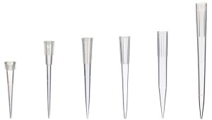
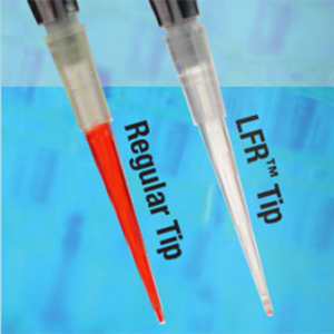
GoldenGate Bioscience universal-fitting pipette tips, are engineered to fit practically all popular manufacturers of single and multichannel pipettes, so reducing the number of tip brands required in a laboratory. Both regular and filter barrier tips are available in GoldenGate’s Low Fluid Retention (LFR™) option.

GoldenGates LFR plasticware is the result over 10 years of research and development in resin technology. This, together with its traditional diamond polished, ultra smooth mold surfaces, accuracy and reproducibility for pipetting liquid in microvolumes is maximised, offering superior pipette tip performance.  In addition, through its optimal fluid transfer, LFR products provide maximum recovery of precious samples and reagents.