Category Archives: Blogging

Field-Deployable Diagnostics: Stability and Performance of Veterinary Rapid Tests Under Variable Environmental Conditions

Veterinary diagnostics are increasingly moving from laboratory-based testing to field-deployable platforms. Among these, lateral flow assays (LFAs) and other rapid immunoassays have become indispensable for detecting pathogens directly in farms, wildlife monitoring sites, and remote veterinary stations. These tools offer speed, portability, and minimal infrastructure requirements, but their performance is strongly influenced by environmental stability factors such as temperature, humidity, transport conditions, and reagent integrity.

This article explores in detail the technical performance considerations, stability validation protocols, and environmental stress impacts of veterinary rapid tests, supported by references to university laboratories, government guidelines, and standardization frameworks.

AffiVET® Canine Brucella Antibody Rapid Test Kit

1. Why Stability Matters in Field-Deployable Veterinary Rapid Tests

Rapid tests in veterinary applications must maintain diagnostic accuracy (sensitivity, specificity, reproducibility) across diverse environments. Unlike centralized laboratories where temperature and humidity are controlled, veterinary field tests are often exposed to:

  • High ambient heat (>40 °C in tropical zones)

  • Cold chain interruptions in shipping and storage

  • Relative humidity fluctuations (10–90 %)

  • Freeze–thaw stress cycles during transport

  • Mechanical shocks during field deployment

Government and academic studies show that environmental instability can result in false negatives, weak bands, or test failure. For example, the USDA APHIS Veterinary Services emphasizes that diagnostic integrity must be protected through packaging and transport stability (USDA APHIS VS).

Similarly, veterinary diagnostic laboratories such as the University of Minnesota VDL (UMN VDL) and Iowa State University VDL (Iowa State VDL) highlight strict shipping protocols because specimen condition directly impacts test outcomes.

2. Environmental Stressors Affecting Veterinary Rapid Tests

2.1 Temperature Fluctuations

  • High temperatures accelerate degradation of antibodies, conjugates, and membrane substrates.

  • Low temperatures slow capillary action, leading to delayed flow and weak or smeared signal bands.

  • Cyclic exposure between hot and cold environments causes polymer fatigue in test housings and irreversible reagent damage.

Reference: Stability testing under accelerated aging protocols is outlined by CLSI EP25-A standards (CLSI Guidelines via FDA).

2.2 Humidity Effects

  • High humidity (>60 % RH) promotes nitrocellulose swelling, leading to inconsistent flow.

  • Moisture can prematurely dissolve conjugates in the pad, resulting in absent control lines.

  • Inclusion of desiccant sachets and barrier foil pouches is recommended.

Academic reviews confirm that humidity management is critical in LFAs (NCBI PMC Lateral Flow Review).

2.3 Freeze–Thaw Stress

  • Each freeze–thaw cycle causes protein denaturation, aggregate formation, and loss of antigen-binding activity.

  • Veterinary samples (e.g., serum, plasma, feces) are particularly prone to degradation, leading to invalid assays.

  • ≤1 freeze–thaw cycle is recommended by veterinary testing laboratories such as UC Davis Veterinary Lab (UC Davis VDL).

2.4 Transport and Mechanical Stress

  • Vibration and mechanical impact during shipping can redistribute conjugate pads or damage cartridge seals.

  • Packaging guidelines from UN3373 Biological Substance regulations (regulated under U.S. Department of Transportation) specify triple packaging for diagnostic specimens (DOT Packaging Regulations).

3. Laboratory and Regulatory Guidelines for Veterinary Rapid Test Validation

Field-deployable diagnostics must comply with performance and stability criteria:

  • USDA APHIS: transport and testing procedures for animal health (USDA Animal Health).

  • CDC Point-of-Care Guidance: lessons from human rapid tests adapted for veterinary applications (CDC POC Testing).

  • University VDLs: institutional protocols from UMN VDL, Iowa State VDL, and UC Davis Veterinary Diagnostics serve as technical references.

These institutions stress:

  • Use of insulated coolers with frozen ice packs.

  • Packaging with leak-proof primary containers.

  • Avoiding direct contact between samples and ice packs to prevent freeze damage.

4. Technical Strategies for Improving Stability

4.1 Advanced Conjugate Stabilization

  • Addition of sugars (trehalose, sucrose) to conjugates to prevent protein unfolding.

  • Use of polymeric stabilizers and lyophilization for long-term room temperature stability.

4.2 Packaging Innovations

  • Aluminum foil pouches with high-barrier properties.

  • Integrated desiccants to reduce RH during storage.

4.3 Thermal Contrast Amplification

  • Embedding metal nanoparticles that enhance signal intensity under variable heat conditions.

  • Proven to improve limit of detection (LOD) by up to 10-fold in veterinary LFAs.

4.4 Rugged Housing Design

  • ABS polymer casings with temperature resistance and UV protection for outdoor deployment.

  • Sealed devices that reduce moisture ingress.

Veterinary rapid test validation under environmental stress is typically organized into stress-testing matrices:

Condition Test Focus Performance Parameter Reference
High Temp (40 °C, 2 weeks) Complete test kit Sensitivity & control band intensity FDA Guidelines
High Humidity (70 % RH, 1 month) Membrane & conjugate pad Flow consistency, false positive rates USDA APHIS
Freeze–Thaw (3 cycles) Antibody conjugates Affinity loss, signal reduction UC Davis VDL
Transport Shock (1 m drop test) Cartridge housing Physical integrity, leakage risk DOT Regulations
Shelf-Life (12–24 months) All reagents Stability profile, accelerated aging prediction CLSI EP25-A

AffiVET® Canine Brucella Antibody Rapid Test Kit

. Conclusion

Veterinary rapid tests deployed in field environments must be rigorously validated for stability and robustness against temperature, humidity, freeze–thaw cycles, and transport disruptions. Governmental agencies like USDA APHIS, as well as leading academic veterinary diagnostic laboratories such as UMN, Iowa State, and UC Davis, provide practical frameworks to ensure test reliability.

By integrating advanced stabilizers, robust packaging, and innovative thermal amplification techniques, manufacturers can deliver rapid tests that remain accurate, reproducible, and durable—making them highly suitable for on-site veterinary diagnostics worldwide.

  • veterinary rapid diagnostic tests

  • lateral flow veterinary test kits

  • field-deployable veterinary diagnostics

  • environmental stability of rapid assays

  • humidity and temperature effects on veterinary diagnostics

  • freeze–thaw stability veterinary assays

  • veterinary diagnostic packaging and transport

  • USDA APHIS veterinary testing protocols

  • university veterinary diagnostic laboratory shipping guidelines

  • accelerated aging CLSI EP25 veterinary diagnostics

PCR External Quality Control: Ensuring Precision and Reliability in Molecular Diagnostics

Polymerase Chain Reaction (PCR) is one of the most powerful tools in molecular diagnostics, revolutionizing the detection of genetic material across numerous fields, from medical diagnostics to forensic analysis. With its vast application range, from detecting pathogens to identifying genetic disorders, ensuring the accuracy and reliability of PCR results is of paramount importance. One critical aspect of maintaining PCR quality is through External Quality Control (EQC), a process that plays a significant role in validating the performance of PCR assays.

What is PCR External Quality Control?

External Quality Control in PCR refers to the practice of verifying the accuracy, consistency, and reproducibility of PCR assays by comparing the results obtained from one laboratory against those from an external reference or control source. Unlike internal controls, which are built into the assay itself, external controls are independent and provide an unbiased check on the assay’s performance. These controls help laboratories ensure that their results are accurate, reproducible, and compliant with regulatory standards.

For example, the CDC often uses external controls in its disease surveillance programs to validate laboratory results across multiple sites. The purpose of EQC is to ensure that laboratory tests can reliably detect the target genetic material and produce reproducible results.

The Importance of External Quality Control in PCR Testing

  1. Ensuring Consistent and Accurate Results
    The main role of external quality control in PCR is to ensure that test results are both consistent and accurate. Using external controls allows laboratories to compare their results against a known, standardized reference, which can help in detecting issues like contamination, poor sample handling, or suboptimal reagent quality. The FDA recommends the use of external controls in PCR-based diagnostics to maintain the highest level of precision and reliability.

  2. Verification of Test Sensitivity and Specificity
    PCR assays must be sensitive enough to detect low concentrations of the target nucleic acids and specific enough to differentiate the target from other genetic material. External quality control samples, often composed of spiked DNA or RNA, allow for the assessment of the assay’s sensitivity and specificity. This is particularly important for detecting rare genetic markers or pathogens. For instance, the NIH suggests that EQC programs should include sensitivity and specificity testing to ensure PCR assays are performing optimally.

  3. Compliance with Regulatory Requirements
    Regulatory bodies like the FDA and the WHO mandate the use of external quality control for diagnostic tests. Regular participation in external quality control programs ensures that laboratories are compliant with these regulatory standards and helps them achieve necessary certifications. External quality control also aids in meeting Good Laboratory Practice (GLP) and Good Manufacturing Practice (GMP) standards, which are essential for obtaining approvals for PCR-based diagnostic kits.

  4. Enhancing Laboratory Confidence
    EQC helps enhance laboratory confidence in their PCR assays by providing an independent verification of the results. By comparing internal results with external benchmarks, laboratories can identify potential biases or errors in their testing process. This allows for early intervention and troubleshooting. The CDC emphasizes that confidence in diagnostic tests is critical, especially when results influence treatment decisions or epidemiological monitoring.

How External Quality Control Works in PCR

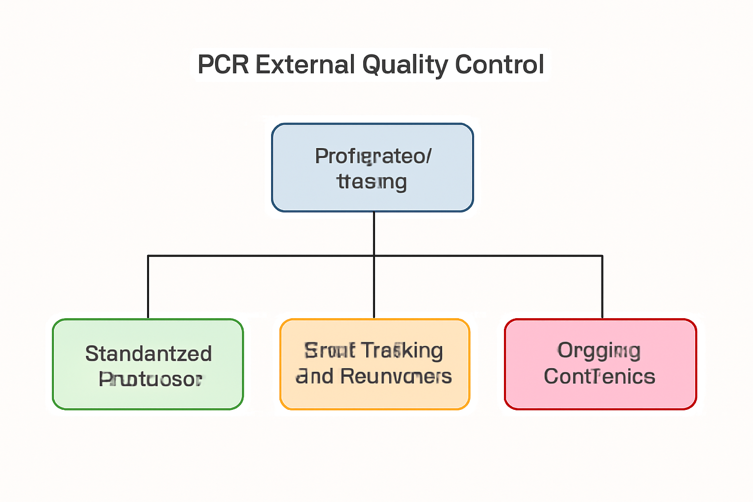
External Quality Control is typically carried out through inter-laboratory comparison programs or proficiency testing. The following steps illustrate how EQC is implemented:

  1. Provision of Blind Samples
    In proficiency testing programs, laboratories receive samples from an external organization, often in a blinded format, meaning the laboratory is unaware of the contents. These samples are designed to mimic real-world specimens and can include both negative and positive controls. The laboratory performs its standard PCR protocol and reports the results back to the external agency.

  2. Comparison with Benchmark Data
    The results from different laboratories are compared against a reference or benchmark data set. This comparison helps to assess the consistency and accuracy of the PCR assay. In many cases, the WHO provides the necessary reference standards for specific diagnostic tests, such as those used for infectious diseases.

  3. Evaluation of Performance
    The external agency evaluates the performance of each laboratory based on how closely their results match the benchmark data. Laboratories that consistently perform poorly may be required to undergo corrective actions, such as improving their assay protocols or recalibrating their equipment.

  4. Feedback and Corrective Action
    Laboratories receive feedback on their performance and are informed if any discrepancies were identified. This feedback is crucial for continuous improvement. The FDA and NIH both stress the importance of incorporating feedback from proficiency testing programs to identify areas for improvement and maintain the quality of PCR-based assays.

Components of a Successful PCR External Quality Control Program

  1. Regular Participation in Proficiency Testing
    Proficiency testing is the cornerstone of an external quality control program. Laboratories should regularly participate in inter-laboratory comparison programs to evaluate their PCR assays’ performance. Programs are often organized by regulatory bodies like the FDA or private entities that specialize in proficiency testing. Regular participation ensures that laboratories stay up-to-date with the latest diagnostic standards.

  2. Standard Operating Procedures (SOPs)
    To ensure the effectiveness of EQC, laboratories must have well-established standard operating procedures (SOPs) in place for their PCR assays. These procedures should be regularly reviewed and updated to ensure they align with best practices and regulatory guidelines. NIST offers resources on developing and maintaining SOPs for molecular diagnostics.

  3. Training and Calibration
    Laboratory staff must be adequately trained to handle proficiency testing samples and interpret results correctly. Routine calibration of PCR equipment, such as thermocyclers, is essential to maintaining assay performance. The CDC recommends regular calibration as part of a quality control program to ensure accurate results.

  4. Error Detection and Root Cause Analysis
    An essential part of EQC is identifying the root causes of any discrepancies between the laboratory’s results and the reference standards. Error detection can involve investigating factors such as reagent quality, sample contamination, or equipment malfunctions. The FDA and NIH both highlight the need for comprehensive error-tracking and resolution procedures to ensure that issues are promptly addressed.

Benefits of External Quality Control for PCR

  1. Improved Test Reliability
    External quality control ensures that PCR assays consistently deliver reliable results. By validating the testing process with external controls, laboratories can avoid errors that might otherwise go unnoticed. The CDC explains that high-quality PCR testing is crucial for disease surveillance and epidemiological studies.

  2. Early Identification of Systemic Issues
    EQC helps detect problems early, before they lead to widespread errors. Whether it’s due to reagent quality, equipment malfunction, or laboratory technique, early identification ensures that corrective actions can be taken swiftly. The NIH underscores the importance of early error detection in maintaining high-quality diagnostic testing.

  3. Ensures Regulatory Compliance
    Regular participation in external quality control programs helps laboratories stay compliant with local and international regulations. Regulatory bodies like the FDA and WHO require evidence of external quality control participation as part of their certification and accreditation processes.

Challenges in Implementing PCR External Quality Control

  1. High Costs
    One of the primary challenges with PCR external quality control is the cost. Participating in proficiency testing and purchasing external controls can be expensive, particularly for smaller laboratories. However, programs offered by organizations like the WHO and CDC may offer financial assistance or lower-cost options for laboratories in low-resource settings.

  2. Standardization Issues
    Despite efforts to standardize external quality control practices, variations in sample handling, reagent preparation, and testing protocols can still lead to discrepancies in results. The NIST is actively working to address these issues through the development of universal standards for molecular diagnostics.

  3. Data Management
    Managing the data generated from proficiency testing programs can be complex, especially for laboratories that participate in multiple EQC programs. Laboratories need robust data management systems to track their performance over time and ensure that corrective actions are taken when needed.

Conclusion

External Quality Control is essential to ensuring the accuracy, reliability, and reproducibility of PCR assays. It allows laboratories to meet regulatory standards, detect issues early, and improve test performance. By implementing proficiency testing, standard operating procedures, and continuous calibration, laboratories can enhance their diagnostic capabilities and ensure they produce consistent, high-quality PCR results.

For more information on PCR quality control and best practices, visit resources from the NIH, FDA, and CDC.

GelRed (10,000X): A Comprehensive Guide to DNA Staining in Molecular Biology

GelRed (10,000X) is one of the most widely used DNA and RNA staining dyes in molecular biology. As a highly sensitive fluorescent stain, it is often used for detecting nucleic acids in gel electrophoresis and other analytical techniques. GelRed offers exceptional performance with high specificity, low toxicity, and versatility across a range of molecular biology applications.

What Is GelRed (10,000X)?

GelRed (10,000X) is a concentrated nucleic acid stain that offers superior fluorescence compared to traditional stains like ethidium bromide. Its high sensitivity enables the detection of even the most minute quantities of DNA or RNA, making it invaluable for precise gel electrophoresis and nucleic acid quantification. The dye binds to nucleic acid molecules through intercalation, producing a bright red fluorescence when exposed to UV light. GelRed is often used in the preparation of agarose gels, polyacrylamide gels, and various other forms of electrophoresis systems.

Unlike ethidium bromide, GelRed is less toxic, reducing potential health hazards in laboratory environments. It also offers improved signal-to-noise ratios, enhancing the clarity of gel bands.

GelRed (10,000X) Properties and Features

High Sensitivity

One of the most significant features of GelRed is its exceptional sensitivity. At 10,000X concentration, it provides a highly concentrated solution that allows users to achieve superior detection sensitivity even with low-concentration DNA. Researchers can visualize minute DNA samples, making it easier to work with very low quantities of nucleic acids for downstream applications such as cloning, quantitative PCR (qPCR), and gene expression analysis.

Compatibility with Various Gels

GelRed is compatible with a range of gel systems, including agarose gels, polyacrylamide gels, and other electrophoresis-based methods. This versatility allows researchers to use GelRed in a wide array of experimental setups, including routine PCR analysis, restriction fragment length polymorphism (RFLP), and Southern blotting. Additionally, GelRed works effectively with high-performance gel systems, such as deep-well agarose gels for specialized applications like quantitative gel electrophoresis.

Low Toxicity

GelRed is designed to be a safer alternative to ethidium bromide, which is known for its potential mutagenic effects and toxicity. GelRed exhibits significantly lower toxicity compared to ethidium bromide and has been found to be non-cytotoxic at working concentrations, which makes it a safer choice for researchers working with high concentrations of nucleic acids.

Source: NIH, Study on GelRed’s Reduced Toxicity

Simple to Use

Unlike traditional dyes that need to be incorporated into the gel prior to electrophoresis, GelRed offers flexibility in its application. The dye can be added directly to the gel or the running buffer, providing researchers with a convenient way to stain DNA and RNA without the need for pre-staining.

Better Resolution

GelRed’s enhanced fluorescence helps in achieving better resolution in electrophoresis experiments, especially when working with complex samples. The dye offers sharp band imaging and lower background noise, which is crucial for accurate molecular analysis and quantification.

Key Applications of GelRed (10,000X)

GelRed (10,000X) is indispensable in several molecular biology applications. Below are the primary uses:

1. PCR Product Visualization

Polymerase Chain Reaction (PCR) is a fundamental technique used to amplify DNA sequences. After amplification, the PCR products must be analyzed to confirm the presence of the target DNA. GelRed is used to stain the gel, allowing for the visualization of PCR bands under UV light. The dye’s high sensitivity allows the detection of low-concentration PCR products with minimal background interference.

Reference: Source on PCR Applications

2. DNA Quantification

GelRed (10,000X) plays a significant role in DNA quantification. Using fluorescence-based methods, GelRed allows researchers to measure the concentration of DNA in a sample. This is crucial in applications such as next-generation sequencing (NGS), where precise DNA quantification is required to ensure that libraries are created with an optimal amount of input DNA.

3. Restriction Fragment Length Polymorphism (RFLP) Analysis

GelRed is also used in restriction fragment length polymorphism (RFLP) analysis, a technique for studying genetic variation. In RFLP, DNA is digested with specific restriction enzymes, and the resulting fragments are visualized using gel electrophoresis. GelRed’s high sensitivity allows for accurate detection of even small differences in fragment length, aiding in genetic mapping and mutation detection.

4. Southern Blotting

Southern blotting is a technique used to detect specific DNA sequences within a complex mixture. After DNA is transferred to a membrane, it is hybridized with a labeled probe, and the presence of the target sequence is detected. GelRed can be used to visualize the DNA bands post-blotting, providing enhanced sensitivity compared to other stains.

5. Genomic Studies

GelRed (10,000X) is used in genomic studies to examine the structure and function of genes. It enables the visualization of genomic DNA after restriction enzyme digestion and electrophoresis. The dye’s minimal toxicity makes it a good option for experiments that require handling large amounts of DNA.

Advantages of GelRed Over Ethidium Bromide

  • Lower Environmental Impact: GelRed can be safely disposed of through regular waste protocols, while ethidium bromide requires special disposal procedures due to its toxicity.

  • Reduced Risk of Mutagenesis: Unlike ethidium bromide, GelRed is not classified as a mutagen, making it safer for long-term laboratory use.

  • Improved Detection: GelRed produces brighter bands and lower background fluorescence, which improves the signal-to-noise ratio for more accurate results.

Handling and Safety Considerations

Although GelRed (10,000X) is less toxic than ethidium bromide, it is essential to follow good laboratory practices. Researchers should wear appropriate protective gear, including gloves and lab coats, when handling the dye. Additionally, all waste containing GelRed should be disposed of properly to minimize environmental impact.

Safety Resources: NIH Safety Information on GelRed

Protocol for Using GelRed (10,000X)

  1. Gel Preparation: Add GelRed to the agarose or polyacrylamide gel at a final concentration of 1X. For a 100 mL gel, add 10 μL of GelRed (10,000X).

  2. Electrophoresis: Run the gel with the sample, and once complete, place the gel under a UV transilluminator.

  3. Imaging: Use a gel documentation system to visualize and capture the fluorescence emitted by the DNA.

Conclusion

GelRed (10,000X) offers exceptional sensitivity, low toxicity, and versatility, making it an indispensable tool in molecular biology research. Its ability to stain DNA and RNA for gel electrophoresis, restriction mapping, and quantification has revolutionized the way molecular biologists approach nucleic acid analysis. Whether used in PCR, Southern blotting, or genomic studies, GelRed ensures precise, reproducible, and safe results for a wide variety of applications.

As the field of molecular biology continues to evolve, GelRed remains a reliable and essential reagent, advancing research in genomics, diagnostics, and beyond.

Comprehensive Overview of RNase Inhibitor ELISA Kit: A Critical Tool for RNA Research

Introduction: The Importance of RNase Inhibitors in RNA-Based Applications

The RNase Inhibitor ELISA Kit is an essential diagnostic tool in the field of molecular biology, offering a highly sensitive and specific method for detecting and quantifying RNase inhibitors. These proteins are vital for preserving RNA integrity in biological samples, making this kit an indispensable tool in a wide array of research applications, including RNA extraction, RT-PCR, and RNA sequencing.

RNase inhibitors play a crucial role in preventing the degradation of RNA by RNases, which are ubiquitous enzymes found in nearly all biological systems. Their application ensures that RNA remains stable and intact, even during rigorous experimental processes. For instance, RNA integrity is fundamental to the accuracy of downstream applications, including quantitative PCR (qPCR) and RNA-Seq, which are used for gene expression studies and biomarker discovery. This kit has been optimized to address the growing demand for accurate and reproducible measurements of RNase inhibitors in diverse experimental setups.

What Is an RNase Inhibitor and Why Is It Important?

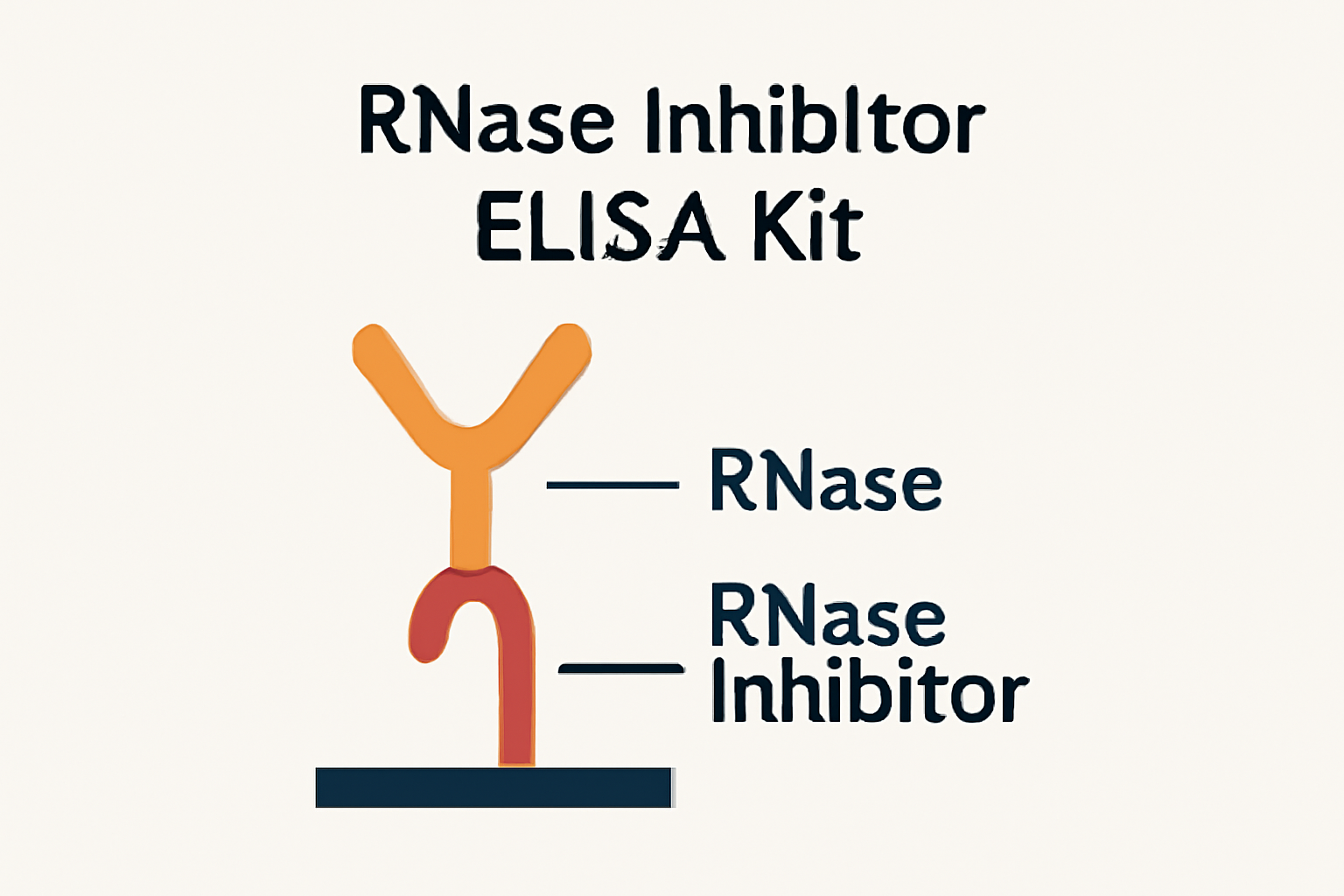
RNase inhibitors are specialized proteins that bind to and neutralize RNases, thus preventing the degradation of RNA molecules. They are typically used in experiments that involve RNA manipulation, where the integrity of the RNA is critical for accurate results. Without RNase inhibitors, RNA molecules can be rapidly degraded, compromising the integrity of experiments.

The Role of RNase Inhibitors in RNA Research

RNA plays a crucial role in many biological processes, and the degradation of RNA by RNases is a significant challenge in molecular biology. Therefore, the use of RNase inhibitors is essential for the following:

  1. RNA Extraction: During the extraction of RNA from biological samples, RNases are often released, which can degrade the RNA if not inhibited. RNase inhibitors prevent this degradation, ensuring high-quality RNA for downstream applications.

  2. RT-PCR: Reverse transcription polymerase chain reaction (RT-PCR) relies on intact RNA templates. RNase inhibitors ensure the reverse transcription step proceeds without the risk of RNA degradation.

  3. RNA-Seq: In RNA sequencing, RNA degradation can result in inaccurate gene expression profiles. RNase inhibitors are routinely used to maintain RNA integrity throughout the entire sequencing process.

For more in-depth information on RNA research and the role of RNases, visit resources such as NIH’s Guide on RNA Extraction and Stanford’s RNA-Seq Overview.

Technical Overview of the RNase Inhibitor ELISA Kit

How the ELISA Kit Works

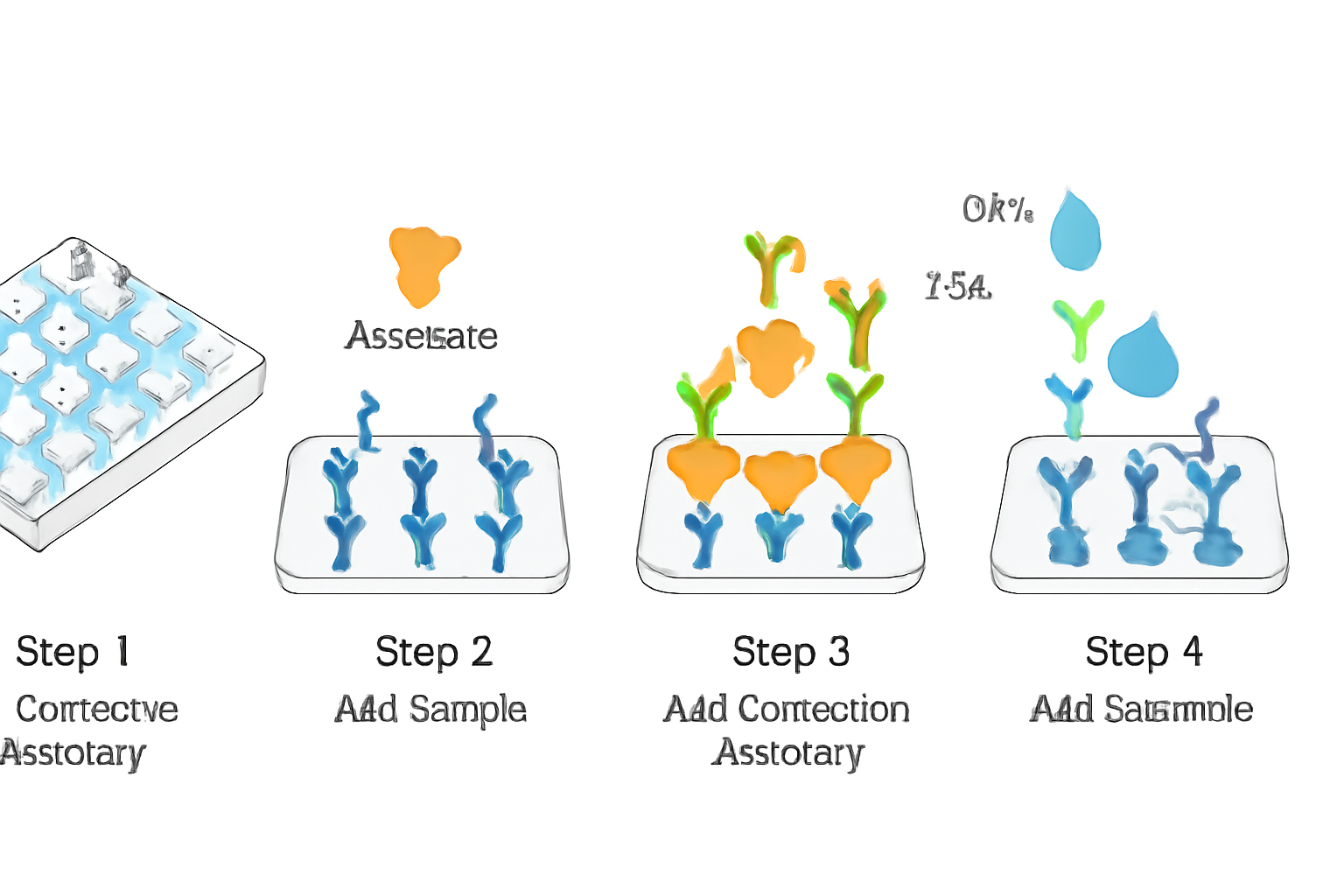
The RNase Inhibitor ELISA Kit employs a sandwich ELISA format, which involves the use of two specific antibodies to detect and quantify RNase inhibitors in biological samples. The kit works by binding the RNase inhibitors to a pre-coated microplate that contains capture antibodies specific to RNase inhibitors. After incubation, the bound inhibitors are detected using an enzyme-conjugated detection antibody that generates a colorimetric signal. The intensity of the color produced is directly proportional to the amount of RNase inhibitor present in the sample.

Key Features of the Kit

  • High Sensitivity: Capable of detecting RNase inhibitors as low as 10 pg/mL.

  • Wide Range of Sample Types: Works with serum, plasma, cell culture supernatants, and tissue lysates.

  • Fast and Simple Protocol: Results can be obtained within 3 hours, making it ideal for high-throughput analysis.

  • Validated in Multiple Platforms: Compatible with 96-well plates and automated systems for large-scale applications.

For information on the general principles of ELISA, visit the FDA ELISA Manual.

AffiELISA® RNase Inhibitor ELISA Kit

Applications of the RNase Inhibitor ELISA Kit

1. RNA Integrity in RNA Extraction

RNase inhibitors are crucial during RNA isolation to prevent degradation by RNases. The RNase Inhibitor ELISA Kit is used to validate the effectiveness of RNase inhibitors in RNA extraction kits. Researchers at Stanford University and NCBI recommend using RNase inhibitors to ensure high-quality RNA for downstream applications like RT-PCR and RNA-Seq.

2. Recombinant Protein Production

In the field of recombinant biotechnology, RNase inhibitors are commonly used to stabilize RNA in in vitro transcription reactions. By quantifying the activity of RNase inhibitors, the kit provides insight into the purity and activity of recombinant proteins used in pharmaceutical and diagnostic applications. Learn more about protein production at NCBI Biotechnology.

3. Gene Expression Analysis

For gene expression studies involving RNA, RNase inhibitors prevent RNA degradation during RT-PCR and RNA-Seq processes. This ensures accurate amplification of mRNA for gene expression profiling. Check out the NIH guide on gene expression analysis.

Sensitivity and Limit of Detection (LOD)

Standard Curve Example

A typical standard curve for the RNase Inhibitor ELISA Kit includes a dilution series of a known RNase inhibitor standard. The relationship between the concentration (pg/mL) and the optical density (OD450) is plotted to generate a linear curve with an R² value of 0.99 or higher. This ensures that even low concentrations of RNase inhibitors can be accurately measured.

For a more detailed explanation of standard curves in ELISA assays, refer to the NIH ELISA Protocols.

Application in RNA Quality Control for RNA-Seq

Role in RNA Sequencing

In RNA sequencing applications, the stability of RNA is paramount. RNase inhibitors are added to RNA samples to prevent degradation before sequencing. This ensures that the RNA is preserved in its original form, leading to more accurate sequencing results.

The RNase Inhibitor ELISA Kit is used in RNA quality control to verify the amount of RNase inhibitor present during RNA extraction, ensuring the protection of RNA through the sequencing process. More about RNA-Seq can be found at NCBI RNA-Seq.

Troubleshooting Guide for RNase Inhibitor ELISA Kit

Problem Cause Solution
High background signal Incomplete washing or excess antibody Increase washing time, reduce antibody concentration
Low signal Inadequate sample preparation Ensure correct sample volume and concentration
Inconsistent results Contamination or improper reagent storage Recheck storage conditions and validate reagent batches

For additional troubleshooting tips, consult the FDA Bioanalytical Method Validation Guide.

Compliance and Safety

  • ISO 13485 Certified: Ensuring consistency in performance and quality.

  • FDA and CE Compliance: Compliant with regulatory requirements for clinical research.

For information on regulatory standards for laboratory kits, visit the FDA’s Biotech Guidance.

Conclusion

The RNase Inhibitor ELISA Kit is an essential tool for researchers working with RNA, providing a reliable and reproducible method for detecting and quantifying RNase inhibitors. Its application in RNA extraction, recombinant protein production, RNA sequencing, and gene expression studies makes it indispensable in molecular biology research.

For more detailed information on the RNase Inhibitor ELISA Kit, including ordering instructions and technical support, visit the following links:

he Role of Leishman Stain in Clinical Hematology and Diagnostics

Overview of Leishman Stain

Leishman Stain is a classic Romanowsky-type stain, primarily used in hematology to examine blood smears. Developed by Sir William Boog Leishman in 1901, the stain is known for its ability to differentiate and highlight the structure of blood cells, enabling pathologists to diagnose a range of hematological disorders. The stain is essential for visualizing various cell types, including red blood cells (RBCs), white blood cells (WBCs), and platelets, each with distinct coloration and structure.

Leishman Stain operates by utilizing a combination of two dyes, methylene blue and eosin, which together allow for a detailed view of cellular components. It is widely utilized not only in clinical settings but also in research and teaching to facilitate the understanding of blood cell morphology. (CDC.gov) (PubMed.ncbi.nlm.nih.gov)

Composition and Chemical Properties

Leishman Stain is a combination of two key components: eosin and methylene blue, both of which contribute to the distinct coloration of blood cells. Methylene blue, a basic dye, stains acidic components, particularly the nuclei of cells, in shades of blue. Eosin, an acidic dye, stains the cytoplasm of cells and other structures like erythrocytes, in varying shades of pink to red. These two components are dissolved in methanol, which not only dissolves the dyes but also acts as a fixative, preparing the blood smear for examination under a microscope.

  • Methylene Blue: This basic dye stains acidic cellular components such as nuclei and granules in cells like neutrophils and basophils. (NIH.gov)

  • Eosin: An acidic dye that stains the cytoplasm, giving cells like RBCs a characteristic pink hue. (NCBI.nlm.nih.gov)

Staining Procedure

Leishman Stain is widely favored due to its straightforward, cost-effective procedure, which can be completed in a relatively short period of time. The steps for preparing and staining a blood smear are as follows:

  1. Preparation of the Blood Smear:

    • Place a drop of blood on a clean glass microscope slide.

    • Use another slide to spread the blood, creating a thin, even smear.

    • Allow the smear to air dry for a few minutes, ensuring the blood is fixed by the methanol in the stain.

  2. Staining the Smear:

    • Apply undiluted Leishman Stain directly onto the dried smear and let it rest for 2–3 minutes.

    • Add an equal volume of distilled water or PBS (phosphate-buffered saline) to the stain, and mix gently. This dilution allows the stain to interact with the blood cells, producing vivid color contrasts.

    • Allow the smear to incubate with the stain and water for 10–15 minutes.

  3. Rinsing and Drying:

    • After the incubation period, rinse the slide gently with PBS or distilled water to remove excess stain.

    • Let the slide dry naturally in a tilted position. Avoid blotting as this may disturb the smear.

  4. Mounting:

    • Once the smear is dry, cover the slide with a mounting medium and place a coverslip on top. The slide is now ready for examination.

This staining process allows for the differentiation of cell types and the identification of abnormalities within the blood smear. (Nature.com) (Labmedicine.com)

Cellular Differentiation and Microscopic Examination

Once stained, the blood smear can be examined under a microscope. The key to Leishman Stain’s effectiveness lies in its ability to highlight the different cellular components of blood, including red blood cells, white blood cells, and platelets. The following features are visible under the microscope:

  • Red Blood Cells (RBCs): These cells appear pink with a central pallor. The size and shape of RBCs can indicate various types of anemia or other blood disorders. (JAMA Network) (Bloodjournal.org)

  • White Blood Cells (WBCs): The WBCs are classified based on their morphological characteristics, including the presence of granules, nucleus shape, and cytoplasm color:

    • Neutrophils: Appear with bluish-pink cytoplasm and multi-lobed nuclei.

    • Lymphocytes: Exhibit a round nucleus surrounded by a thin rim of blue cytoplasm.

    • Monocytes: Display a kidney-shaped nucleus and gray-blue cytoplasm.

    • Eosinophils: Characterized by orange-red cytoplasmic granules.

    • Basophils: Present with dark purple cytoplasmic granules.

  • Platelets: Platelets appear as small, purple cytoplasmic fragments, vital in the evaluation of thrombocytopenia or thrombocytosis. (PubMed.ncbi.nlm.nih.gov) (NIH.gov)

AffiSTAIN® Leishman Stain

Clinical Applications of Leishman Stain

Leishman Stain plays a critical role in diagnosing various blood-related conditions. Its ability to differentiate between different cell types allows clinicians to assess blood smears for various disorders, including:

  • Anemia: By evaluating the size, shape, and color of RBCs, Leishman Stain can help identify different types of anemia, such as microcytic anemia (small RBCs) and macrocytic anemia (large RBCs).

  • Leukemia: Leishman Stain is useful for identifying abnormal white blood cells, such as blasts in acute leukemia or characteristic cell types in chronic leukemia.

  • Infectious Diseases: Leishman Stain can be used to detect parasites such as Plasmodium (malaria) and Trypanosoma (sleeping sickness) in blood samples, which are often crucial for the diagnosis of these diseases. (CDC.gov) (WHO.int)

  • Platelet Disorders: The stain helps assess platelet count and morphology, essential in diagnosing conditions like thrombocytopenia or thrombocytosis. (NIH.gov) (NCBI.nlm.nih.gov)

Advantages of Leishman Stain

Leishman Stain is preferred in many laboratory settings due to several advantages:

  • Simplicity: The staining procedure is straightforward and does not require sophisticated equipment. This makes it accessible to various laboratories, especially in low-resource settings.

  • Cost-Effective: Compared to other staining methods like Wright’s Stain or Giemsa Stain, Leishman Stain is relatively inexpensive, which makes it an excellent option for routine hematological analysis.

  • Speed: The staining procedure can be completed relatively quickly, allowing for rapid results in diagnostic settings.

Limitations of Leishman Stain

Despite its widespread use, Leishman Stain has a few limitations:

  • Resolution: While effective for general blood smear examination, Leishman Stain may not offer the resolution needed for more advanced diagnostic techniques like cytogenetic analysis or molecular studies.

  • Stability: The working solution of Leishman Stain can be less stable and may require frequent preparation, which could be a limitation in high-throughput laboratories.

  • Specificity: For certain diseases or conditions, additional staining methods, such as immunohistochemistry, may be required to identify specific cell markers or pathogens more accurately.

Conclusion

Leishman Stain remains a vital and indispensable tool in hematology and clinical diagnostics. Its ability to differentiate various blood cells with a straightforward, cost-effective, and rapid procedure makes it a cornerstone in the diagnostic process. While there are limitations regarding its resolution and specificity, Leishman Stain continues to be a reliable and valuable technique in clinical laboratories worldwide. For more in-depth protocols and research, consider exploring educational and governmental resources like the National Institutes of Health (NIH) and the Centers for Disease Control and Prevention (CDC).

Additional Resources: For further reading, you can visit the following authoritative resources:

Comprehensive Guide to Colorimetric Assay Kits and Inorganic Pyrophosphatase Assays

Introduction to Colorimetric Assays

Colorimetric assays are essential tools in biochemical and molecular research for quantifying various biomolecules, including proteins, nucleic acids, and enzymes. These assays are based on a color change triggered by the interaction between a substrate and an enzyme, allowing researchers to measure the activity of enzymes or other molecules present in the sample. This simple yet highly effective method is used across a wide range of disciplines, from molecular biology to clinical diagnostics.

The significance of colorimetric assays is demonstrated in their widespread application in biological and chemical analysis. Colorimetric assays can provide valuable insights into various metabolic and enzymatic processes by translating complex biochemical reactions into measurable color changes, making them one of the most versatile methods in modern biochemistry.

For further reading on colorimetric methods, see the NIH article on colorimetric assay applications.

Inorganic Pyrophosphatase Assays: Understanding the Role of IPPase

Inorganic pyrophosphatase (IPPase) is a vital enzyme involved in cellular metabolism. It catalyzes the hydrolysis of inorganic pyrophosphate (PPi) into two phosphate molecules, releasing energy that is essential for various cellular functions. IPPase plays a crucial role in regulating energy balance and supporting many fundamental biological processes, including DNA replication, protein synthesis, and cellular signaling.

IPPase activity can be monitored using colorimetric assays, which have become a standard tool for measuring enzyme activity. These assays are particularly useful in research focused on metabolic pathways, energy production, and the study of diseases that disrupt cellular energy regulation.

Explore more about IPPase and its biological importance in this PubMed article on the role of IPPase in cellular metabolism.

Principle of Colorimetric Assay Kits

Colorimetric assay kits are designed to detect enzymatic reactions through the production of a color change, which is proportional to the amount of enzyme or substrate present in a sample. These kits typically include specific reagents, buffers, and substrates that are optimized for the detection of specific enzyme activities, such as IPPase.

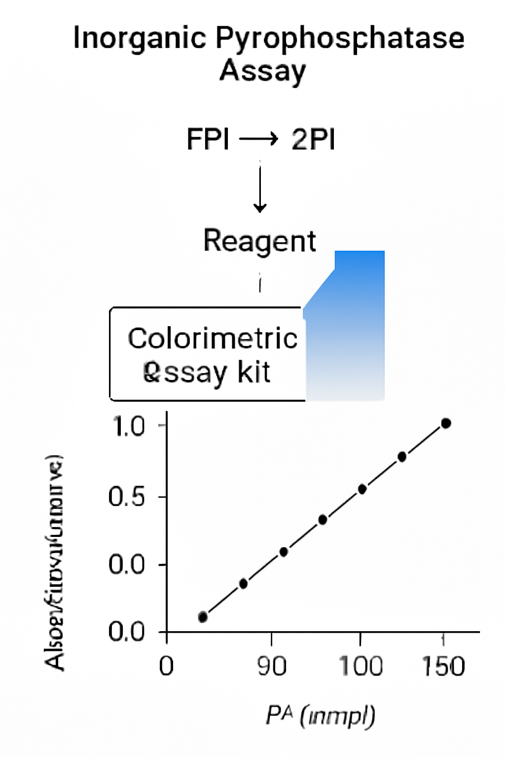
In a typical colorimetric assay for IPPase, the reaction involves the hydrolysis of inorganic pyrophosphate, leading to the production of free phosphate ions. The phosphate ions react with a dye or substrate that produces a visible color change, which can be measured using a spectrophotometer. The intensity of the color is directly proportional to the enzyme’s activity, providing quantitative data on enzyme concentration.

For more details on assay principles, visit NIH’s resource on assay techniques.

Applications of Colorimetric Assays in Research

Colorimetric assays are incredibly versatile, with applications in various fields of scientific research, including:

1. Energy Metabolism Research

Understanding energy metabolism is central to many biological studies. Colorimetric assays that measure IPPase activity can provide insights into how cells manage energy resources. By quantifying the activity of enzymes like IPPase, researchers can gain a deeper understanding of cellular energy regulation and how metabolic processes are altered in diseases such as cancer, diabetes, and neurological disorders.

To learn more about energy metabolism and IPPase’s role in it, visit PubMed – IPPase and Energy Metabolism.

AffiASSAY® MTS Cell Proliferation Colorimetric Assay Kit

2. Biochemical Pathway Analysis

Colorimetric assays allow scientists to track the activity of enzymes and metabolites involved in biochemical pathways. These assays are used in metabolic flux analysis, where the flow of metabolites through various enzymatic reactions is measured. In the case of IPPase, these assays are valuable for studying its involvement in pathways related to phosphate metabolism and energy production.

Further insights into biochemical pathways can be found in NCBI – Biochemical Pathways.

3. Drug Discovery and Screening

The colorimetric assay kit for IPPase is an essential tool in drug discovery, particularly for screening inhibitors or activators of the enzyme. By assessing the impact of potential drug candidates on IPPase activity, researchers can identify promising molecules that modulate this enzyme’s function. These assays are invaluable for discovering treatments for diseases caused by metabolic dysregulation.

For more on drug screening methods, visit NIH – Drug Screening Methods.

Advantages of Colorimetric Assays

Colorimetric assays offer several distinct advantages, which make them a preferred choice for researchers in many fields:

  • Sensitivity: These assays are highly sensitive, allowing for the detection of even minute quantities of enzyme activity.

  • Simplicity: The protocols for colorimetric assays are straightforward and do not require complex instrumentation, making them accessible in most laboratories.

  • Quantification: The color change provides quantitative data, which can be analyzed to determine enzyme activity levels accurately.

  • Versatility: They can be used for a wide range of enzymes and biomolecules, including IPPase, making them suitable for various experimental applications.

  • High Throughput: Colorimetric assays can be adapted to high-throughput screening, enabling large-scale studies on enzyme activity.

For a more detailed comparison of assay types, refer to EPA – Assay Methodology.

Technical Considerations for Successful Assays

While colorimetric assays are straightforward, certain technical considerations must be taken into account to ensure accurate and reproducible results:

1. pH and Buffer Optimization

The pH of the reaction mixture is critical for enzyme stability and activity. IPPase, like most enzymes, has an optimal pH range in which it exhibits the highest activity. Using the correct buffer composition is essential for maintaining the enzyme’s activity during the assay. Refer to NIH – Buffer Optimization for further guidelines.

2. Incubation Time

The reaction time must be optimized to ensure that the color change is detectable without being too prolonged. Over-incubation can lead to over-saturation, while under-incubation may result in weak signals. Proper timing is crucial to obtain accurate results.

3. Calibration Standards

Using calibration standards is essential for converting absorbance readings into enzyme activity units. It’s important to include known concentrations of phosphate to establish a standard curve, allowing the conversion of absorbance into enzyme concentration.

Innovative Advancements in Colorimetric Assays

The field of colorimetric assays continues to evolve with advancements in assay sensitivity and automation. Some recent innovations include:

  • Microfluidic Devices: The integration of microfluidic technology into colorimetric assays allows for faster and more efficient analysis with reduced reagent volumes.

  • Enhanced Reagents: New reagents with improved sensitivity are being developed, allowing for the detection of even lower enzyme concentrations.

  • Automation: High-throughput automation systems are being incorporated into colorimetric assays, enabling large-scale studies with minimal human intervention.

For further reading on the advancements in assay technologies, check PubMed – Recent Developments in Assays.

Case Studies in IPPase Research

Research on IPPase is critical for understanding how cells manage their energy reserves. One area where IPPase assays have proven valuable is in the study of bone metabolism. IPPase has been shown to regulate osteoclast differentiation, and colorimetric assays have been used to study its role in bone resorption.

Furthermore, colorimetric assays are used in research focused on neurological disorders, where IPPase dysregulation can affect cellular energy homeostasis. Studies measuring IPPase activity in neuronal cells have provided insights into its role in neurodegeneration.

Explore case studies on IPPase in bone metabolism in this PubMed article on osteoclast differentiation.

Conclusion: The Future of Colorimetric Assays in Biochemistry

Colorimetric assays for IPPase and other enzymes have revolutionized biochemical research by offering a simple yet effective method for quantifying enzyme activity. As the technology behind these assays continues to evolve, we can expect even more accurate, sensitive, and efficient methods for measuring enzyme function. The continued development of these assays will be crucial in advancing our understanding of metabolic processes and drug discovery, particularly in areas related to energy metabolism.

Stay up to date with advancements in colorimetric assays through this NIH resource on assay techniques.


References

  1. NIH – Colorimetric Assays

  2. PubMed – Pyrophosphate and Cellular Energy

  3. NIH – Assay Techniques

  4. PubMed – Role of IPPase in Energy Metabolism

  5. EPA – Assay Standardization

Comprehensive Guide to Human Inorganic Pyrophosphatase ELISA and Inorganic Pyrophosphatase Assays

Introduction to Inorganic Pyrophosphatase (IPPase)

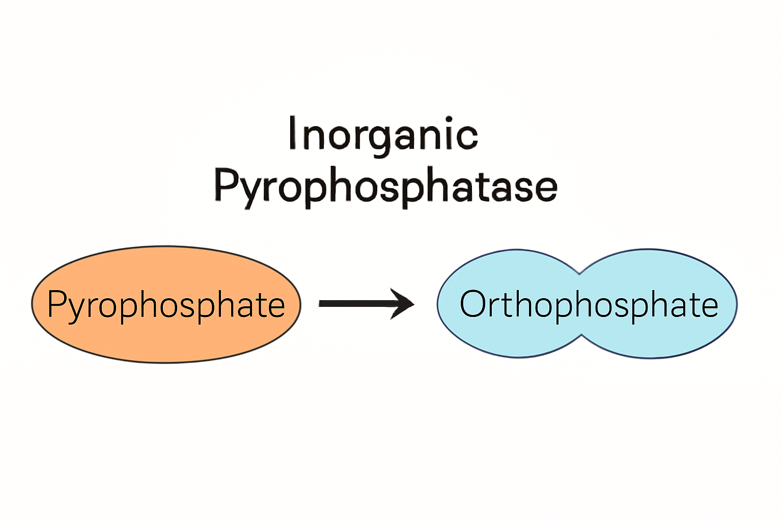
Inorganic pyrophosphatase (IPPase, EC 3.6.1.1) is an essential enzyme involved in the hydrolysis of inorganic pyrophosphate (PPi) to orthophosphate (Pi). This reaction is vital for maintaining the balance between ATP and inorganic phosphate in cells, playing a key role in nucleotide metabolism and energy regulation (NCBI Gene). IPPase is found in a variety of cellular compartments, including the cytosol, mitochondria, and plastids, and its activity is tightly regulated due to its involvement in critical cellular functions.

Enzyme Function and Significance

The primary function of IPPase is to catalyze the breakdown of PPi, a byproduct of several biosynthetic reactions, into two molecules of Pi. This reaction provides cellular energy by removing PPi, thus driving forward the biochemical processes that generate ATP. The hydrolysis of PPi is particularly important in DNA synthesis, RNA transcription, protein synthesis, and various metabolic pathways.

Given its critical function, abnormalities in IPPase activity have been implicated in various diseases and metabolic disorders. Therefore, accurately measuring IPPase levels in biological samples is crucial for understanding these pathologies.

The Human Inorganic Pyrophosphatase ELISA Kit

The Human Inorganic Pyrophosphatase ELISA Kit is a highly sensitive immunoassay designed for the detection and quantification of human IPPase in different biological samples, including serum, plasma, tissue extracts, and cell lysates. This kit offers researchers a reliable tool to monitor IPPase activity and investigate its role in human health and disease.

ELISA Principle and Mechanism

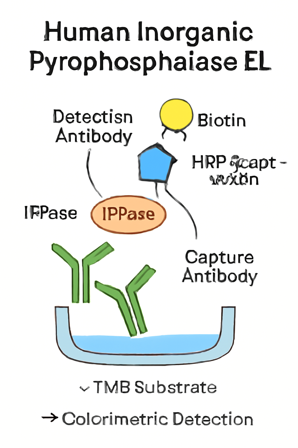
The ELISA (Enzyme-Linked Immunosorbent Assay) principle used in the Human IPPase ELISA Kit is based on the sandwich immunoassay technique, where two antibodies are used to capture and detect the target antigen. The basic steps are as follows:

  1. Coating of the microplate: The plate is pre-coated with an anti-IPPase capture antibody.

  2. Sample incubation: The biological sample containing the target IPPase is added to the wells and binds to the capture antibody.

  3. Detection: A biotinylated secondary antibody specific to IPPase is added, followed by streptavidin-HRP, which reacts with the substrate to produce a detectable color change.

  4. Measurement: The intensity of the color change is proportional to the amount of IPPase in the sample and is measured at 450 nm.

This method is widely used because of its sensitivity, specificity, and ability to provide quantitative results. It is especially useful in clinical diagnostics and research settings where precise measurement of IPPase levels is necessary.

For further reading on ELISA techniques, consult NCBI ELISA Protocols.

Applications of Inorganic Pyrophosphatase Measurement

The quantification of Inorganic Pyrophosphatase (IPPase) is essential for understanding various biological processes and diseases. Here are some of the key areas where IPPase plays a significant role:

1. Cancer Metabolism

In cancer, metabolic reprogramming is a hallmark of tumor cells. A study from the National Cancer Institute emphasizes the role of pyrophosphate metabolism in tumor progression. IPPase is involved in maintaining cellular ATP levels and facilitating nucleic acid biosynthesis, processes that are altered in cancers. Quantifying IPPase activity can provide insights into cancer cell metabolic shifts and may aid in developing targeted therapies.

2. Neurological Disorders

Neurodegenerative diseases, including Alzheimer’s disease and Parkinson’s disease, often involve disrupted cellular homeostasis and energy regulation. According to PubMed, abnormalities in pyrophosphate metabolism are associated with neuronal dysfunction. IPPase assays are used in neurology research to study how IPPase activity influences neuronal health and to assess potential therapeutic targets.

3. Mitochondrial Function and Energy Homeostasis

Mitochondria are the energy powerhouses of the cell, and their function is tightly linked to the regulation of ATP and inorganic phosphate. The NIH Mitochondrial Disease Research program highlights the importance of maintaining proper pyrophosphate levels in mitochondrial metabolism. Alterations in IPPase activity can lead to mitochondrial dysfunction, which is a hallmark of many mitochondrial diseases.

4. Metabolic Disorders

The disruption of phosphate metabolism and energy production often leads to metabolic disorders such as glycogen storage diseases and mucopolysaccharidoses. The NIH Genetic and Rare Diseases Information Center (GARD) provides extensive information on how enzymes like IPPase contribute to the regulation of metabolic pathways and how these pathways can be exploited for therapeutic purposes.

AffiELISA® Human Inorganic pyrophosphatase ELISA [ PPA1]

Inorganic Pyrophosphatase Assay Techniques

Beyond ELISA kits, enzymatic assays provide another method for quantifying IPPase activity in biological samples. These assays typically measure the release of inorganic phosphate (Pi) upon the hydrolysis of PPi. The two most common techniques for this type of analysis include:

1. Molybdate-based Phosphate Assays

These assays are based on the reaction between phosphate and molybdate, which forms a phosphomolybdate complex. The intensity of this complex can be measured spectrophotometrically at 620 nm. This method is widely used in biochemical research for quantifying Pi released during enzymatic reactions.

For additional assay protocols, see the PubChem Assay Protocols.

2. Colorimetric Assays Using Coupled Enzyme Reactions

Another method for measuring IPPase activity involves coupling the PPi hydrolysis reaction with a secondary enzyme system. This can amplify the signal for better detection. These assays are often used when higher sensitivity or lower concentrations of IPPase are involved.

Quality Control and Validation of Assays

Assay quality is critical for obtaining reproducible and accurate results. The following steps are essential in ensuring that IPPase assays, whether ELISA or enzymatic, are properly validated:

  • Intra-assay Precision: The coefficient of variation (CV) should be < 10% for consistent results across replicate samples.

  • Inter-assay Precision: CV across multiple runs should be < 15% to ensure assay reproducibility.

  • Linearity of Dilution: A standard curve should show a linear relationship across the concentration range, with a recovery rate between 95–105%.

  • Spike Recovery: The addition of known amounts of IPPase should result in a recovery rate of 90–110%, confirming the assay’s accuracy.

For guidelines on bioanalytical method validation, refer to the FDA’s Validation Guidelines.

Troubleshooting IPPase Assays

Even with high-quality kits, researchers may encounter problems during the assay process. Below are common issues and troubleshooting strategies:

Problem Possible Cause Solution
High background Insufficient washing Increase washing time or number of washes
Low signal Incomplete antibody binding Ensure proper incubation times and temperatures
Inconsistent results Plate shaker malfunction Calibrate or replace shaker to ensure consistent mixing

For detailed troubleshooting, check the NIH Assay Validation Guide.

Future Directions in IPPase Research

The future of IPPase research looks promising, particularly with the advancements in high-throughput screening technologies, which allow for large-scale studies on enzyme function. Some potential areas of focus include:

  • Gene editing techniques, such as CRISPR-Cas9, to investigate the role of IPPase in specific diseases.

  • The development of specific inhibitors targeting IPPase to treat metabolic disorders and cancer.

  • The use of IPPase inhibitors in conjunction with other metabolic modulators to assess synergistic effects on cell growth and survival.

As our understanding of IPPase deepens, these research areas will continue to evolve, potentially leading to new therapeutic strategies for metabolic diseases and cancer.

Conclusion

The Human Inorganic Pyrophosphatase ELISA Kit and Inorganic Pyrophosphatase Assays are indispensable tools in biological and medical research. These assays provide vital insights into the role of IPPase in cellular metabolism, energy regulation, and disease. By utilizing these advanced techniques, researchers can gain deeper insights into pyrophosphate metabolism, offering potential therapeutic avenues for metabolic disorders, cancer, and neurodegenerative diseases.

For more information and updates on IPPase research, please refer to trusted sources such as PubMed, National Institutes of Health (NIH), and Gene Ontology Consortium.

Streptavidin Magnetic Beads: Enhancing Biomolecule Isolation and Purification

Streptavidin magnetic beads have transformed the way biomolecules are isolated and purified, offering a highly efficient and versatile method for use in various laboratory applications. Streptavidin, a protein that binds biotin with high specificity and affinity, is attached to magnetic beads, creating a tool that allows for easy separation of biotinylated molecules using a magnetic field. The combination of streptavidin’s robust binding properties and the magnetic beads’ separation capabilities has made this system a cornerstone of many biochemical assays, especially in proteomics and genomics.

Fundamentals of Streptavidin-Biotin Binding

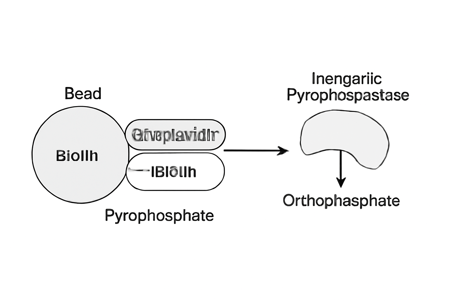
The streptavidin-biotin interaction is one of the strongest non-covalent bonds known in nature. The binding affinity is so high that it is widely used in various laboratory applications to achieve high-purity biomolecule isolation. This interaction forms the basis of numerous assays, from immunoprecipitation to protein purification and DNA/RNA extraction. Biotinylation of molecules, such as antibodies, proteins, or nucleic acids, allows them to be captured by streptavidin-coated magnetic beads. This enables researchers to perform highly efficient and selective isolations.

  • High Binding Affinity: Streptavidin binds biotin with a dissociation constant (Kd) of approximately 10^(-15) M, making it ideal for sensitive applications such as quantitative PCR or mass spectrometry.

  • Magnetic Separation: The inclusion of magnetic beads allows for the rapid separation of biomolecules from complex mixtures, simplifying sample preparation and reducing handling times. This is especially advantageous in high-throughput experiments where speed and efficiency are critical (NCBI – Streptavidin-Biotin Interaction).

Applications in Protein Purification and Immunoassays

One of the most common uses of streptavidin magnetic beads is in protein purification. Biotinylated proteins or antibodies can be selectively captured by streptavidin-coated beads, facilitating their isolation from complex biological mixtures. This is particularly useful in immunoassays, such as Western blotting, immunohistochemistry, and ELISA.

Protein-Protein Interaction Studies

Streptavidin magnetic beads are also used in protein-protein interaction studies, a critical area of research in drug discovery. By capturing biotinylated interacting partners, researchers can examine complex networks of protein interactions. This has far-reaching implications in studying signal transduction pathways, cellular responses to drugs, and post-translational modifications.

Link to Research:

Nucleic Acid Purification and Detection

In addition to protein isolation, streptavidin magnetic beads are highly effective for nucleic acid purification, especially in the context of DNA and RNA extraction. Biotinylated oligonucleotides or primers are used to capture complementary nucleic acid sequences, providing a highly specific method for purification.

  • DNA/RNA Extraction: The beads allow for the isolation of genomic DNA or RNA from complex biological samples, facilitating downstream applications like quantitative PCR or next-generation sequencing (NGS). This is particularly valuable in clinical diagnostics and genetic research.

  • Nucleic Acid Hybridization Assays: Streptavidin magnetic beads are used in hybridization assays to detect specific nucleic acid sequences, such as in Southern or Northern blotting, enabling the study of gene expression and mutation detection.

Link to Research:

AffiBEADS® Streptavidin Magnetic Beads

Inorganic Pyrophosphatase Assays: A Key Tool for Metabolic Studies

Inorganic pyrophosphatases (PPases) are enzymes that hydrolyze inorganic pyrophosphate (PPi) into two phosphate ions. These enzymes play a fundamental role in cellular metabolism, driving various biosynthetic reactions by providing energy through the hydrolysis of pyrophosphate. In biochemical research, inorganic pyrophosphatase assays are used to study the activity of PPases, investigate enzyme kinetics, and develop therapeutic strategies targeting pyrophosphate metabolism.

Biological Significance of Inorganic Pyrophosphatases

Inorganic pyrophosphatases are critical for the biosynthesis of nucleotides and the regulation of energy transfer in cells. They help maintain cellular ATP levels by catalyzing the breakdown of pyrophosphate, which otherwise could accumulate and inhibit essential metabolic processes. PPases are found in nearly all living organisms, from bacteria to humans, and are involved in numerous biological processes.

  • Metabolic Pathways: PPases contribute to the synthesis of nucleic acids, lipids, and carbohydrates. Their action is particularly important in the formation of ATP during DNA replication, RNA transcription, and protein synthesis.

  • Energy Regulation: PPases help maintain energy balance in cells by regulating pyrophosphate levels, a key factor in metabolic control. The hydrolysis of pyrophosphate provides energy for many biosynthetic processes, ensuring proper cellular function.

Applications of Inorganic Pyrophosphatase Assays in Drug Discovery

Inorganic pyrophosphatase assays are widely used in drug discovery to investigate enzyme kinetics and substrate specificity. These assays help identify potential inhibitors of pyrophosphate metabolism, which can serve as leads for new therapeutic agents.

Enzyme Kinetics and Inhibition Studies

By studying the kinetics of inorganic pyrophosphatases, researchers can determine key parameters such as the Michaelis-Menten constant (Km) and the maximum reaction velocity (Vmax). These measurements are essential for understanding the enzyme’s role in metabolic processes and for identifying potential inhibitors that could modulate its activity. PPase inhibitors are being explored as therapeutic agents in cancer therapy, where metabolic regulation is critical for tumor growth, and in antimicrobial treatments, where targeting pyrophosphate metabolism may reduce bacterial resistance.

  • Enzyme Inhibition: Inorganic pyrophosphatase inhibitors have been shown to modulate metabolic pathways, and the development of specific inhibitors has potential for treating various diseases, including cancer and bacterial infections.

Link to Research:

Inorganic Pyrophosphatase in Cancer and Microbial Research

The role of inorganic pyrophosphatase in regulating cellular energy makes it an important target for cancer research. Tumor cells often exhibit altered metabolic pathways to support uncontrolled growth, and PPases play a role in maintaining this metabolic shift. Inhibition of PPases may disrupt the energy supply required for tumor survival.

Moreover, PPase inhibitors are being tested as potential antibiotics to combat drug-resistant bacteria. By targeting the pyrophosphate metabolism in microbes, PPase inhibitors could reduce bacterial resistance to existing treatments, providing a new avenue for antimicrobial drug development.

Link to Research:

Conclusion: Advancements in Streptavidin Magnetic Beads and Inorganic Pyrophosphatase Assays

Both streptavidin magnetic beads and inorganic pyrophosphatase assays represent powerful tools in biochemical and molecular biology research. Streptavidin magnetic beads have revolutionized biomolecule isolation by providing a rapid, efficient, and scalable method for protein, DNA, and RNA purification. Their applications in immunoassays, protein-protein interaction studies, and nucleic acid isolation have significantly advanced our understanding of cellular processes.

Similarly, inorganic pyrophosphatase assays offer critical insights into cellular energy metabolism, providing the foundation for drug discovery and therapeutic interventions targeting metabolic diseases, including cancer and microbial infections. As these technologies evolve, they will continue to shape the future of molecular research and therapeutics, driving innovations in diagnostics and treatment strategies.

References:

Tissue Total RNA Isolation Kit: Comprehensive Overview for High-Quality RNA Extraction

Total RNA isolation is a critical step in molecular research, enabling scientists to study gene expression, molecular pathways, and regulatory mechanisms at the RNA level. The Tissue Total RNA Isolation Kit is designed to efficiently extract RNA from diverse tissue types, ensuring optimal RNA quality for downstream applications such as RNA sequencing, qPCR, and microarray analysis.

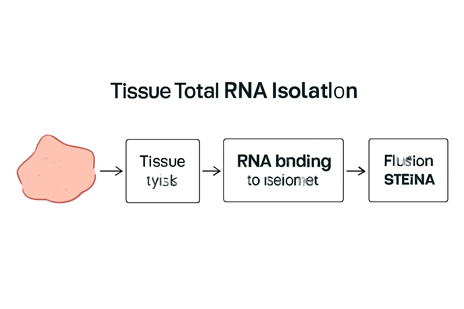
RNA molecules are delicate and susceptible to degradation by ribonucleases (RNases), which are ubiquitous in the environment. For this reason, RNA isolation protocols must be highly optimized to prevent RNA degradation during extraction. The Tissue Total RNA Isolation Kit achieves this by utilizing a combination of specialized lysis buffers and a proprietary column-based purification method. This process ensures the isolation of high-purity RNA that is free from contaminants, allowing for accurate gene expression analysis.

Key Features of the Tissue Total RNA Isolation Kit:

  • High yield of intact RNA from challenging tissue samples, including those with low RNA content.

  • Fast and efficient protocol with minimal hands-on time.

  • Low RNase contamination, facilitated by the use of RNase inhibitors in the lysis buffer.

  • Compatibility with a variety of downstream applications, such as RNA-Seq, microarray analysis, and quantitative PCR (qPCR).

Optimizing RNA Quality for Accurate Data

The RNA integrity number (RIN) is a measure of RNA quality, and it directly influences the success of downstream analyses. A high RIN value indicates the presence of full-length, intact RNA molecules, which are essential for accurate transcriptomic analyses. When isolating RNA, the most common contaminations to avoid include genomic DNA (gDNA), proteins, and lipids, all of which can negatively affect the purity and integrity of the RNA. These contaminants are often removed during the RNA extraction process, ensuring that only high-quality RNA is isolated for use in downstream assays.

When using the Tissue Total RNA Isolation Kit, researchers benefit from the optimized buffer system that ensures efficient RNA isolation while preventing contamination from genomic DNA. This is particularly important for RNA sequencing experiments, where even low amounts of DNA contamination can significantly impact data quality.

For comprehensive guidelines and tips on RNA extraction, you can visit resources like PubMed Central (PMC) and NCBI Gene Expression (NCBI Gene Expression) for detailed protocols and troubleshooting advice.


The Role of RNA in Cellular and Molecular Research

RNA plays a crucial role in translating genetic information stored in DNA into functional proteins that carry out the cellular functions essential for life. There are various types of RNA, each with specific functions:

  • Messenger RNA (mRNA): Carries genetic information from DNA to the ribosome for protein synthesis.

  • Ribosomal RNA (rRNA): Forms the core structure of the ribosome and catalyzes protein synthesis.

  • Transfer RNA (tRNA): Helps in the translation of mRNA into proteins by transporting amino acids to the ribosome.

One of the most significant uses of RNA isolation is the study of differential gene expression. Researchers can compare mRNA expression levels between different tissues or conditions, enabling the identification of genes that are upregulated or downregulated in response to specific biological processes or diseases. By isolating RNA from various tissue samples, including tumor tissues, scientists can explore the molecular mechanisms underlying diseases such as cancer, neurodegeneration, and metabolic disorders.

For example, cancer research has heavily relied on RNA isolation techniques to identify cancer biomarkers. By analyzing RNA expression profiles from healthy versus diseased tissues, scientists can pinpoint key genes associated with tumor progression. Further details on RNA sequencing in cancer research can be explored through resources like Cancer.gov (NIH Cancer).

AffiPURE®  Cell/Tissue Total RNA Isolation Kit

Applications of RNA Isolation in Disease Research

RNA isolation plays a pivotal role in understanding various diseases, particularly those with genetic underpinnings. A few notable areas of research include:

  • Cancer genomics: By isolating RNA from cancerous tissues, scientists can study how tumor cells hijack normal cellular processes, leading to uncontrolled growth.

  • Neurological diseases: RNA profiling from brain tissues can help identify genes involved in neurodegenerative diseases such as Alzheimer’s and Parkinson’s.

  • Infectious diseases: RNA analysis can also shed light on how viruses such as HIV, HCV, and SARS-CoV-2 hijack host cellular machinery for replication.

Through RNA sequencing (RNA-Seq) and other profiling methods, these applications advance our understanding of disease mechanisms and provide insights into potential therapeutic targets. Learn more about these applications in journals published by the National Institutes of Health (NIH) and Centers for Disease Control and Prevention (CDC).


Inorganic Pyrophosphatase Assays: Measuring Key Enzyme Activity in Metabolic Pathways

Inorganic pyrophosphatase (PPase) is an essential enzyme that catalyzes the hydrolysis of inorganic pyrophosphate (PPi) into inorganic phosphate (Pi). This reaction is pivotal in cellular metabolism, as PPi accumulation can inhibit critical metabolic processes, including DNA replication, protein synthesis, and lipid biosynthesis.

PPase activity is important in maintaining cellular energy balance, as it helps in controlling the levels of pyrophosphate within cells. Elevated PPi levels can disrupt metabolic processes, leading to cellular dysfunction. Therefore, precise measurement of PPase activity is essential for understanding enzyme kinetics and the regulatory mechanisms controlling cellular metabolism.

Inorganic Pyrophosphatase Assay Methods

The Inorganic Pyrophosphatase Assay involves the measurement of inorganic phosphate (Pi) released during the hydrolysis of PPi. This is typically done using colorimetric or fluorometric detection methods. In the colorimetric approach, a color change occurs as Pi reacts with a dye, allowing researchers to quantify the enzyme’s activity. The fluorometric method, on the other hand, uses a fluorescent probe that binds to Pi, providing more sensitive detection of small amounts of Pi.

Both techniques are highly valuable for studying PPase kinetics and characterizing inhibitors or activators of the enzyme. PPase assays are essential for research in metabolic diseases, where dysregulation of pyrophosphate metabolism may be a contributing factor.

Key Applications of Inorganic Pyrophosphatase Assays

  • Metabolic research: Understanding the role of PPase in various metabolic pathways, including ATP synthesis and biosynthesis of nucleotides.

  • Therapeutic research: PPase inhibitors are being explored as potential drugs for diseases such as cancer and neurological disorders.

  • Enzyme kinetics: Studies of PPase kinetics help elucidate the enzyme’s role in cellular energy production.

Researchers can find detailed methodologies and applications of PPase assays in resources from NCBI Bookshelf (NCBI Bookshelf) and PubMed (PubMed).


Conclusion: Integrating RNA Isolation and Enzyme Assays for Advancing Research

The Tissue Total RNA Isolation Kit and Inorganic Pyrophosphatase Assays are indispensable tools for researchers in fields ranging from genomics to metabolic disease. By isolating RNA with high integrity and measuring PPase activity with precision, these products contribute to our understanding of gene expression, enzyme regulation, and cellular metabolism.

These technologies are vital for exploring disease mechanisms, developing therapeutic strategies, and advancing biomedical research. Whether studying cancer, neurological diseases, or metabolic disorders, these tools provide essential insights that help push the boundaries of modern science.

For further exploration of these topics and to access comprehensive protocols, please visit NCBI Gene Expression (NCBI Gene Expression) and PubMed (PubMed) for peer-reviewed research articles.

Comprehensive Guide on RIG-I, RNase Inhibitor, and Inorganic Pyrophosphatase Assays

Introduction

In molecular and cellular biology, precise assays are fundamental tools for studying critical enzymes and proteins like RIG-I, RNase inhibitors, and inorganic pyrophosphatase (PPase). These molecules are central to the functioning of immune responses, RNA preservation, and energy metabolism. Assays that measure their activity and presence are indispensable in advancing research in immunology, molecular genetics, and biochemistry.

This detailed guide delves into the biochemical roles of RIG-I, RNase inhibitors, and PPase, with a focus on the assays designed to detect, quantify, and study these molecules. Additionally, the article provides insights into optimizing these assays and their applications in research.

RIG-I (Retinoic Acid-Inducible Gene I): Overview and Assay Techniques

RIG-I is a cytoplasmic receptor involved in the immune system’s response to viral infections, specifically recognizing double-stranded RNA (dsRNA). It is a key player in the innate immune system and triggers downstream signaling cascades that lead to antiviral responses, including the production of interferons. Given its critical role, precise quantification and functional assessment of RIG-I are crucial in immunology and virology.

Function of RIG-I in Immune Defense

RIG-I functions as a pattern recognition receptor (PRR) and is primarily responsible for detecting viral RNA. Upon recognition of viral RNA, RIG-I undergoes conformational changes that activate downstream pathways involving IRF3 and NF-kB. These pathways lead to the production of type I interferons (IFNs) and other inflammatory cytokines, which help to initiate antiviral responses.

  • Activation Pathways: The recognition of dsRNA triggers the binding of RIG-I to the RNA molecule, followed by conformational changes that lead to its interaction with the mitochondrial antiviral-signaling protein (MAVS). This interaction activates the NF-kB and IRF3 pathways, ultimately resulting in the production of antiviral proteins.

Assay Techniques for RIG-I

Detecting the activity and expression of RIG-I is performed using several assays, each designed to target different aspects of RIG-I activation or expression. The most common assays include:

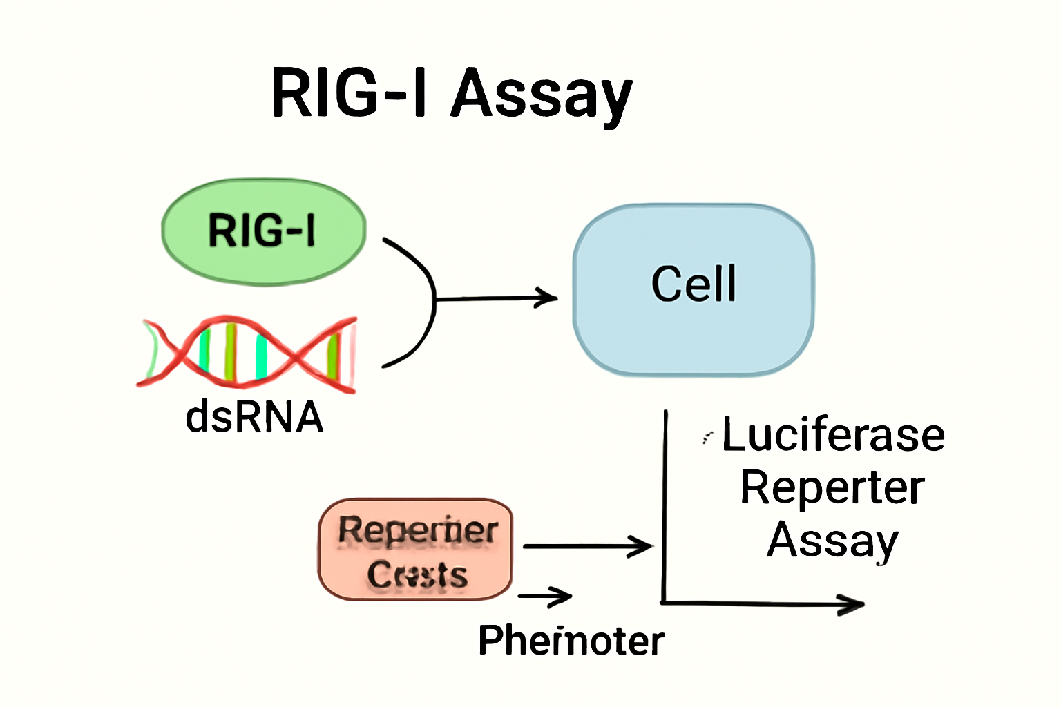
  1. Luciferase Reporter Assay:

  2. Western Blotting:

    • Purpose: Detects RIG-I protein levels.

    • Protocol: Cell lysates are subjected to gel electrophoresis, followed by transfer to a membrane and detection with RIG-I-specific antibodies.

  3. ELISA (Enzyme-Linked Immunosorbent Assay):

    • Purpose: Quantifies RIG-I expression.

    • Protocol: An ELISA kit specific for RIG-I is used to measure its concentration in cell lysates or serum.

  4. qRT-PCR (Quantitative Reverse Transcription PCR):

    • Purpose: Measures RIG-I mRNA expression.

    • Protocol: cDNA synthesis followed by PCR quantifies RIG-I mRNA levels in cells treated with RNA mimics or infected with viruses.

For further detailed exploration on RIG-I activation and its assays:


RNase Inhibitors: Mechanisms and Assays

RNase inhibitors are essential in protecting RNA molecules from degradation during experimental procedures. These inhibitors bind to ribonucleases (RNases), preventing them from degrading RNA during applications such as RT-PCR, RNA sequencing, and RNA isolation.

Mechanism of RNase Inhibition

RNase inhibitors, such as RNasin or RNAseOUT, specifically inhibit the enzymatic activity of RNases by binding to the catalytic site of the RNase enzyme, thus preventing the RNA degradation. These inhibitors are crucial when working with RNA in experiments that require RNA integrity, especially in PCR or RNA purification.

Key Functions:

  • Preservation of RNA Integrity: RNase inhibitors ensure the stability of RNA during extraction and analysis.

  • Prevent RNA Degradation: By binding to RNases, these inhibitors allow researchers to isolate and work with intact RNA for downstream applications.

AffiELISA® RIG-I dsRNA ELISA Kit

Assay Methods for RNase Activity

  1. Fluorogenic Assays:

    • Purpose: To quantify RNase activity.

    • Protocol: RNase activity is detected by measuring the fluorescence emitted when a fluorogenic RNA substrate is cleaved by the RNase enzyme.

    • References:

  2. Gel Electrophoresis:

    • Purpose: To assess RNA degradation.

    • Protocol: RNA samples are subjected to gel electrophoresis, and the integrity is assessed based on the size of the RNA bands. Degradation by RNase results in smaller RNA fragments.

  3. Western Blotting for RNase Inhibitors:

    • Purpose: Detects the presence of RNase inhibitors.

    • Protocol: Protein samples are analyzed by SDS-PAGE followed by Western blotting with RNase inhibitor-specific antibodies.

For further details on RNase inhibitors and their applications:


Inorganic Pyrophosphatase (PPase): Biochemical Role and Assay Methods

Inorganic pyrophosphatase (PPase) catalyzes the hydrolysis of inorganic pyrophosphate (PPi) into inorganic phosphate (Pi), an essential step in energy metabolism. PPase plays a key role in ATP synthesis and hydrolysis, making it a critical enzyme in energy homeostasis within cells.

Function of PPase in Metabolism

The hydrolysis of PPi by PPase is vital in various biochemical processes, including:

  • ATP Synthesis: PPase activity aids in maintaining an equilibrium between ATP and PPi levels.

  • Cellular Energy Homeostasis: PPase regulates the balance of energy within the cell by driving reactions that utilize ATP.

Assay Methods for PPase Activity

  1. Spectrophotometric Assay:

    • Purpose: Measures PPase activity by quantifying inorganic phosphate release.

    • Protocol: The assay uses a spectrophotometric reaction where phosphate is measured using colorimetric detection.

  2. Radiolabeled Substrate Assay:

    • Purpose: Uses 32P-labeled PPi to trace its hydrolysis to Pi.

    • Protocol: The radioactive signal is measured to determine the rate of PPi hydrolysis by PPase.

  3. Fluorometric Assay:

    • Purpose: Quantifies Pi production.

    • Protocol: Fluorescent substrates are used to detect Pi, which is released upon hydrolysis of PPi by PPase.

For further reading on PPase and its assays:


Conclusion

The study of RIG-I, RNase inhibitors, and inorganic pyrophosphatase is central to understanding vital biological processes such as immune responses, RNA preservation, and cellular energy metabolism. The assays developed to measure the activity of these molecules provide critical insights into molecular biology, immunology, and biochemistry.

By employing these assays effectively, researchers can unlock new knowledge about the molecular mechanisms underpinning diseases and therapeutic strategies. For further detailed protocol references, continue exploring these resources: