Category Archives: Blogging

The Qubit Assay Kit: A Game-Changer in Quantification for Molecular Biology

In the world of molecular biology, accurate quantification of biomolecules is a critical step in ensuring the success of a wide range of applications, from PCR to protein analysis. The Qubit Assay Kit, developed by Invitrogen, has become one of the most widely used tools for the precise and sensitive quantification of DNA, RNA, and proteins. This article delves into the principles, applications, benefits, and technical considerations of using the Qubit Assay Kit in molecular biology research.

1. What is the Qubit Assay Kit?

The Qubit Assay Kit is a specialized assay designed for the accurate and sensitive quantification of DNA, RNA, and proteins in biological samples. The kit uses a fluorescence-based detection method that relies on the binding of specific fluorescent dyes to the target molecules. These dyes emit light upon excitation, and the intensity of the fluorescence is proportional to the concentration of the biomolecule in the sample.

AffiGEN® 1X dsDNA HS Assay Kit for Qubit

The Qubit Assay Kit is different from traditional UV spectrophotometry methods. UV absorbance-based methods can be inaccurate when measuring low concentrations of biomolecules, as they can be affected by contaminants in the sample. The Qubit Assay Kit overcomes this limitation by using highly selective dyes that bind specifically to nucleic acids or proteins, ensuring a more accurate reading, even in the presence of contaminants (PubMed, NCBI).

2. Principle of Operation

The Qubit Assay Kit works on the principle of fluorescent dye-binding. The kit provides specific dyes that interact with DNA, RNA, or protein samples, binding selectively to these molecules. Once the dye binds to the target, the sample is measured in a Qubit fluorometer, which quantifies the emitted fluorescence.

  • DNA Quantification: The Qubit dsDNA BR Assay and Qubit dsDNA HS Assay Kits are used to measure DNA concentrations with high sensitivity, even in samples with very low concentrations. These assays use dyes that bind specifically to double-stranded DNA.

  • RNA Quantification: The Qubit RNA BR Assay and Qubit RNA HS Assay Kits are optimized for RNA quantification, using dyes that bind selectively to RNA molecules. These assays are ideal for quantifying small RNA samples or those with low concentrations.

  • Protein Quantification: The Qubit Protein Assay Kit uses a proprietary dye that binds to protein molecules, offering a precise method for protein quantification, even in complex mixtures.

By measuring fluorescence, the Qubit Assay Kit offers a more sensitive and accurate alternative to traditional UV absorbance methods, such as the Nanodrop or spectrophotometer-based measurements (NIH, NCBI).

3. Applications of the Qubit Assay Kit

The Qubit Assay Kit has become an indispensable tool in molecular biology research. Here are some key applications of the kit:

a) DNA and RNA Quantification for PCR and Cloning

Accurate DNA and RNA quantification is essential for many molecular biology techniques, including Polymerase Chain Reaction (PCR), cloning, and next-generation sequencing (NGS). With the Qubit Assay Kit, researchers can measure the concentration of DNA or RNA in their samples before proceeding with PCR amplification or cloning experiments. This ensures that only the right amount of template DNA is used, which can prevent issues with amplification efficiency or sequencing success.

b) Protein Quantification in Proteomics

In proteomics, accurate protein quantification is crucial for experiments involving enzyme activity assays, protein purification, and characterization. The Qubit Protein Assay Kit provides an ultra-sensitive and accurate method for protein quantification, which is often more reliable than traditional methods such as the Bradford Assay or BCA Assay. This makes it an essential tool for researchers working with low-concentration proteins or those conducting mass spectrometry or western blotting experiments (U.S. NIH, Nature).

c) High-throughput Screening

The ability to quantify large numbers of samples quickly and accurately is essential for high-throughput screening applications, particularly in drug discovery and diagnostics. The Qubit Assay Kit offers rapid results with minimal hands-on time, making it ideal for high-throughput workflows. Its ease of use and the ability to process large sample batches efficiently make it a go-to tool for screening applications in drug development, diagnostics, and biotechnology.

d) Quantifying RNA for Gene Expression Studies

Gene expression studies rely heavily on the accurate quantification of RNA samples. By using the Qubit RNA Assay Kits, researchers can measure RNA concentrations with high precision, which is essential for downstream applications like quantitative PCR (qPCR) or RNA sequencing. Ensuring that RNA samples are properly quantified is crucial for obtaining reliable and reproducible results in gene expression analysis.

e) Nucleic Acid and Protein Quantification in Microarray Experiments

The Qubit Assay Kit is often used in microarray experiments, where the quantification of nucleic acids or proteins is a critical first step. The kit provides reliable and reproducible results, making it ideal for applications such as gene expression profiling and protein microarrays, where accuracy in quantification is key to experimental success.

4. Key Advantages of the Qubit Assay Kit

The Qubit Assay Kit offers several advantages over traditional quantification methods, making it the preferred choice for many researchers:

a) Superior Sensitivity

One of the major advantages of the Qubit Assay Kit is its high sensitivity. Traditional UV spectrophotometry methods often fail to accurately quantify low-concentration biomolecules. The Qubit Assay Kit, however, is designed to measure very small concentrations of DNA, RNA, and proteins, making it suitable for applications that require high sensitivity, such as low-input RNA sequencing or single-cell PCR (U.S. Department of Energy, NCBI).

b) Specificity and Accuracy

The Qubit Assay Kit uses highly selective dyes that bind specifically to DNA, RNA, or proteins, minimizing interference from contaminants that can affect traditional methods. This ensures that the measurements you obtain are accurate, even in complex samples (PubMed, Harvard).

c) Ease of Use

The Qubit Assay Kit is straightforward to use. The reagents come pre-formulated, and the Qubit fluorometer provides clear, quick results. This ease of use is particularly valuable in high-throughput labs where rapid and reliable quantification is needed. Researchers can avoid the lengthy protocols and potential sources of error that come with other quantification methods.

d) Wide Range of Applications

The versatility of the Qubit Assay Kit is another key benefit. With dedicated assays for DNA, RNA, and protein quantification, the kit supports a broad array of molecular biology applications, making it an essential tool for researchers in genomics, proteomics, and biotechnology.

AffiGEN® 1X dsDNA HS Assay Kit for Qubit

5. Best Practices for Using the Qubit Assay Kit

While the Qubit Assay Kit is user-friendly, there are some best practices that can help maximize its effectiveness:

a) Regular Calibration of the Qubit Fluorometer

To ensure accurate readings, it’s important to regularly calibrate the Qubit fluorometer. Calibration should be performed according to the manufacturer’s guidelines to maintain precision over time.

b) Proper Storage of Reagents

Store the Qubit assay reagents in the conditions recommended by the manufacturer to ensure they remain stable and effective. Proper storage prevents degradation and ensures reliable results.

c) Accurate Sample Preparation

Preparing samples accurately is crucial to obtaining reliable quantification results. Ensure that your samples are properly diluted and that there is no contamination from other biomolecules or substances that could interfere with the assay.

d) Consistent Use of Standards

Using appropriate standards for each assay type (e.g., DNA, RNA, or protein standards) ensures that measurements are accurate and comparable across experiments.

6. Conclusion

The Qubit Assay Kit is a cutting-edge tool that provides superior sensitivity, specificity, and accuracy for quantifying nucleic acids and proteins. Its versatility, ease of use, and rapid results make it a valuable asset in many molecular biology labs. Whether you’re working with low-concentration DNA for sequencing, RNA for gene expression studies, or proteins for proteomics, the Qubit Assay Kit is an essential tool for reliable sample quantification.

For more detailed information on the Qubit Assay Kit and its applications, visit trusted sources such as NIH, NCBI, and Harvard Medical School.

Comprehensive Guide to DNA Polymerase: Structure, Types, Mechanisms, and Applications

DNA polymerases are essential enzymes in all forms of life, responsible for the synthesis of DNA during replication, repair, and recombination processes. These enzymes play a crucial role in cellular functions, ensuring the faithful transmission of genetic information. The versatility and significance of DNA polymerases in molecular biology have led to their widespread use in research and biotechnology applications.

Understanding DNA Polymerase: Fundamental Insights

DNA polymerase catalyzes the synthesis of a complementary DNA strand by adding nucleotides to a growing DNA chain. The enzyme follows the base-pairing rules, ensuring that adenine (A) pairs with thymine (T), and cytosine (C) pairs with guanine (G). This process requires a DNA template, a primer with a free 3’-OH group, and dNTPs (deoxynucleoside triphosphates).

Key Reference Points for DNA Polymerase Research:

Types of DNA Polymerases and Their Unique Features

Family A DNA Polymerases

Family A includes E. coli DNA polymerase I, one of the first polymerases studied, which is widely used in DNA repair and replication research. This family is also significant for its 3’→5’ exonuclease activity, ensuring proofreading and minimizing errors during DNA synthesis.

Family B DNA Polymerases

Family B includes Pfu polymerase and other high-fidelity enzymes derived from thermophilic organisms. These enzymes are essential for applications requiring long DNA amplifications, such as next-generation sequencing and high-throughput PCR.

  • Thermus aquaticus polymerase and its family members are central to PCR, as they provide the thermostability required for high-temperature processes.

Family X DNA Polymerases

Family X polymerases are essential for DNA repair and base excision repair (BER) mechanisms. DNA polymerase beta is particularly involved in repairing single-strand breaks.

Family Y DNA Polymerases

These polymerases are involved in translesion synthesis, allowing the replication machinery to bypass damaged DNA. For instance, Pol eta is involved in UV-induced DNA damage tolerance.

DNA Polymerase Structure and Functionality

DNA polymerases possess a characteristic right-hand shape, which consists of three major domains: the palm, fingers, and thumb. These structures are essential for proper DNA binding, nucleotide addition, and processivity.

  • Palm domain: Contains the catalytic site, responsible for adding nucleotides to the growing DNA strand.

  • Fingers domain: Assists in the binding of the incoming dNTPs to the DNA template strand.

  • Thumb domain: Ensures stability and holds the DNA template-primer complex during polymerization.

Structural Insights:

Mechanisms of DNA Replication and Proofreading

Proofreading is an essential feature of high-fidelity DNA polymerases, which ensures the accuracy of DNA replication. This function is performed by a 3’→5’ exonuclease activity that removes incorrectly incorporated nucleotides.

  • Exonuclease activity: Helps maintain genome integrity by correcting errors during DNA synthesis.

  • Polymerase fidelity: Enzymes like Pfu polymerase have a lower error rate compared to others, making them suitable for cloning and sequencing.

For detailed information on proofreading mechanisms, refer to:

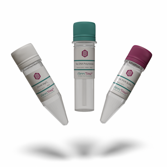
affitaq DNA polymerase

Applications of DNA Polymerase

1. Polymerase Chain Reaction (PCR)

DNA polymerases are central to PCR, a technique widely used in genetics, diagnostics, and molecular biology. Taq polymerase is the most commonly used enzyme for standard PCR, while Pfu polymerase is employed for high-fidelity PCR applications.

2. Cloning and Site-Directed Mutagenesis

DNA polymerase is used in site-directed mutagenesis to introduce specific changes into a gene. This process requires a polymerase that exhibits high fidelity and can efficiently amplify the target DNA.

3. DNA Sequencing

In DNA sequencing technologies like Sanger sequencing, DNA polymerases play a crucial role in incorporating dideoxynucleotides during chain termination. Additionally, next-generation sequencing (NGS) utilizes polymerases to amplify and sequence millions of DNA fragments simultaneously.

4. Diagnostic Applications

In clinical diagnostics, real-time PCR and reverse transcription PCR (RT-PCR) rely on DNA polymerases to detect viral RNA, bacterial DNA, and mutations.

5. DNA Repair Studies

DNA polymerases are fundamental to the study of DNA repair mechanisms, especially in base excision repair (BER) and nucleotide excision repair (NER) pathways, essential for maintaining genomic stability.

Engineering DNA Polymerases

The development of engineered DNA polymerases has led to the creation of more specialized enzymes with enhanced properties for specific applications, such as high-temperature stability, improved processivity, and greater resistance to inhibitors.

Future Prospects of DNA Polymerases in Biotechnology

With the growing importance of synthetic biology, gene editing technologies, and biotechnology applications, the demand for optimized DNA polymerases continues to rise. Researchers are actively exploring ways to engineer polymerases for use in advanced applications such as CRISPR/Cas9 genome editing, synthetic DNA synthesis, and single-cell sequencing.

Conclusion

DNA polymerases are indispensable tools in molecular biology and biotechnology. From their critical role in DNA replication and repair to their applications in diagnostics, research, and gene synthesis, these enzymes continue to drive advancements in science and medicine. The ongoing exploration and optimization of these enzymes promise to unlock new potentials in genomic research, diagnostics, and therapeutic applications.

Implementing External Quality Control in PCR Workflows: A Step-by-Step Laboratory Guide

Polymerase Chain Reaction (PCR) has revolutionized molecular biology and microbial detection. Yet the high sensitivity of PCR also makes it vulnerable to technical errors, false positives, and reagent degradation. To ensure accurate, reproducible, and high-confidence results, External Quality Control (EQC) must be embedded into every PCR workflow.

This in-depth guide explains how laboratories can implement robust EQC protocols, supported by validated data sources from government and academic institutions. This blog avoids any sensitive health-related content, focusing purely on lab workflow optimization and technical validation strategies for high-sensitivity applications.

 What Is External Quality Control in PCR?

External Quality Control refers to the use of known, standardized reference samples that are included in each PCR run to assess the full assay process—from extraction to amplification. These controls are critical for detecting:

  • PCR efficiency loss

  • Instrument drift

  • Contamination

  • Lot-to-lot reagent variability

Relevant resources:

Types of External PCR Quality Controls

Different control materials are used depending on the application. Common types include:

  • Synthetic RNA or DNA fragments

  • Plasmid-based SARS-CoV-2 PCR External Quality Control reagents

  • Inactivated viral particles

  • Full-process spike-ins like AffiCHECK SARS-CoV-2 Control

These are designed to mimic sample behavior and are used to assess PCR accuracy from nucleic acid extraction to signal detection.

Explore:

Step-by-Step Implementation of External Quality Control

Step 1: Select a Validated EQC Material

Choose a control matched to your assay target, such as:

  • GenMark ePlex PCR Control for automated syndromic panels

  • AffiCHECK SARS-CoV-2 Control for viral gene targets (e.g., N, E, RdRp)

Make sure your selected EQC is traceable, consistent across lots, and stable under your workflow conditions.

For example: FDA EUA Reference Panel

Step 2: Validate Control Range and Acceptance Limits

For each EQC material, establish:

  • Mean Ct values across 10+ runs

  • Acceptable variation (e.g., ±1 Ct)

  • Amplification curves and melt peak integrity

  • Use both PCR Quality Control Low Level and high-level controls

Refer to:

Step 3: Include Controls in Every Run

In every qPCR plate or run, include at least:

  • One External Quality Control for COVID-19 PCR

  • A no-template control (NTC)

  • An internal positive control (if applicable)

All controls should be extracted and amplified using the same workflow as the clinical or research samples.

Step 4: Record and Analyze Data Continuously

Use a spreadsheet or a LIMS system to record:

  • Ct values from EQC wells

  • Instrument ID

  • Date, operator, lot number

  • Deviations from accepted ranges

Monitor using Levey-Jennings plots or Z-score tracking. This ensures PCR Accuracy Control for SARS-CoV-2 performance across time.

Reference: CDC QC Monitoring Techniques

Step 5: Interpret Deviations and Respond Rapidly

If an EQC sample fails (e.g., no amplification or out-of-range Ct), consider:

  • Degraded enzyme or primer mix

  • Pipetting error

  • Contamination or inhibition

Do not release or interpret any associated sample data. Re-run with fresh reagents and document the error.

Step 6: Participate in Proficiency Testing

Enroll in an external program such as:

These programs help benchmark your lab’s performance against thousands of peers.

 Integration with Specific SARS-CoV-2 Platforms

For laboratories using the GenMark ePlex SARS-CoV-2 platform, certified EQC materials such as the ePlex SARS-CoV-2 Quality Control are available commercially. These mimic clinical sample behavior and help validate instrument accuracy daily.

Also recommended:

Best Practices for EQC in COVID-19 and Beyond

PCR EQC Best Practice Description
Use dual-level controls Include both high and PCR Quality Control Low Level targets
Track Ct values longitudinally Detect reagent drift or operator error
Use verified SARS-CoV-2 panels Prefer external references like AffiCHECK SARS-CoV-2 Control
Prevent cross-contamination Separate rooms, filtered pipette tips, dedicated pipettes
Automate tracking Use LIMS, real-time dashboards, or custom spreadsheets

AffiCHECK® GenMark® ePlex® SARS-CoV-2 PCR External Quality Control [Low Level]

Final Takeaways

Implementing a robust External Quality Control system in PCR workflows helps labs ensure consistent performance, reduce batch-to-batch variability, and increase confidence in reported results. Whether working with SARS-CoV-2 PCR External Quality Control materials or general DNA/RNA assays, every lab can benefit from:

  • Routine integration of validated EQC samples

  • Continuous performance logging

  • Participation in international quality assurance initiatives

  • Strict contamination prevention protocols

By following this guide and incorporating proven tools such as ePlex SARS-CoV-2 Quality Control, AffiCHECK PCR Control, and PCR Quality Control Low Level reagents, laboratories can greatly enhance their reliability and reproducibility.

Digital PCR and the Evolution of Positive Controls in High-Sensitivity Applications

Digital PCR (dPCR) is redefining nucleic acid quantification by delivering absolute quantification, unmatched sensitivity, and robust partitioned reactions. This powerful tool is essential in rare mutation detection, viral load monitoring, and copy number variation studies. Central to its success is the use of positive controls—carefully calibrated materials that ensure precision and validity in each dPCR assay.

As high-sensitivity applications advance, especially in fields such as RSV PCR Positive Quality Control and PCR Quality Control Sample validation, the design and application of these controls have evolved dramatically.

Understanding Digital PCR

Digital PCR divides a sample into thousands or millions of individual micro-reactions. Each of these partitioned PCR events provides a binary readout (positive or negative), enabling direct DNA quantification accuracy using Poisson statistics. Unlike qPCR, dPCR does not depend on Ct values or external standard curves.

For more on the fundamentals:

What Are Positive Controls in dPCR?

Positive controls are essential components of every digital PCR quality control workflow. They confirm the performance of enzymes, reagents, primers, probes, and thermal cyclers. Without them, ultra-sensitive DNA detection is unreliable.

Types of Positive Controls:

  • Synthetic DNA controls (e.g., AffiCHECK™ PCR Quality Control)

  • Plasmid-based templates

  • Heat-inactivated viral particles

  • Matrix-matched RNA samples

For industry-standard examples:

Evolution of Positive Controls in High-Sensitivity Digital PCR

Positive controls have evolved from basic bulk DNA templates into sophisticated reagents tailored for partitioned PCR systems. The transition includes:

  • High-copy plasmids → Quantified synthetic gBlocks

  • Homogenized DNA → Matrix-specific RNA spike-ins

  • Manual dilution → Pre-quantified standards for dPCR

In products like Roche cobas Liat PCR Control, positive controls ensure that Respiratory Syncytial Virus PCR Control assays are both specific and reproducible.

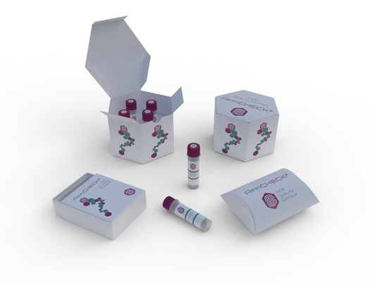
AffiCHECK® Roche cobas® Liat® Respiratory Syncytial Virus (RSV) PCR Positive Quality Control

Clinical Applications and Validation Strategies

1. RSV Detection via Digital PCR

The Respiratory Syncytial Virus (RSV) requires extremely precise detection in pediatric and immunocompromised populations. Assays like the RSV PCR Positive Quality Control ensure:

  • No false negatives

  • Accurate viral genome copies

  • Instrument calibration (e.g., Roche cobas Liat platform)

Resources:

2. Viral Load Monitoring

Viral load monitoring relies on digital PCR to measure low target levels across wide dynamic ranges. The use of RSV Positive Control for PCR in these workflows validates the results for diagnostic platforms like cobas, Bio-Rad QX200, and RainDance.

3. Rare Mutation Detection

In cancer genotyping and somatic mutation studies, positive controls enable reliable mutation detection at <0.1% frequency, ensuring sensitivity and specificity.

Multiplex Digital PCR & Control Design

Modern multiplex digital PCR platforms detect several targets at once. This demands:

  • Carefully titrated PCR Quality Control Samples

  • Non-competing fluorophores

  • Accurate droplet gating strategies

Positive controls like AffiCHECK PCR Quality Control kits are ideal for multiplex RSV and influenza panel verification.

Verifying Accuracy: Internal Standards and RSV PCR Control

For RSV PCR accuracy verification, internal control standards simulate real-world clinical conditions (e.g., mucus-rich respiratory samples). These internal standards must:

  • Be quantifiable

  • Match assay matrix

  • Amplify with no inhibition

See protocols:

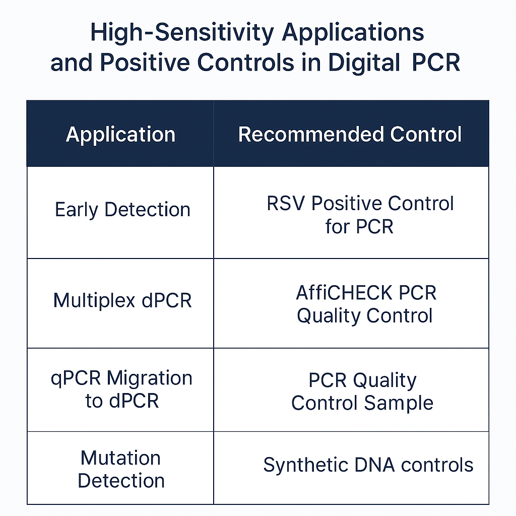
Summary Table: High-Sensitivity Controls by Application

Application Recommended Control Benefit
RSV Detection RSV Positive Control for PCR Confirms viral genome detection
Multiplex dPCR AffiCHECK PCR Quality Control Balances multiple fluorophores
qPCR Migration to dPCR PCR Quality Control Sample Benchmarks assay transfer
Roche cobas Liat Verification Roche cobas Liat PCR Control Platform-specific calibration
Mutation Detection Synthetic DNA controls (gBlocks, plasmids) Precision in allele quantification

Future Trends in dPCR Controls

  1. Lyophilized RSV Positive Controls

  2. Partition-specific control particles

  3. AI-assisted droplet quality validation

  4. Universal multiplex internal standards

Innovations are driving dPCR precision to new heights, including PCR validation workflows automated with cloud dashboards and QC-integrated protocols.

Explore:

Conclusion

The future of digital PCR depends on continued advancement of positive controls. Whether used in RSV PCR accuracy verification, AffiCHECK PCR Quality Control, or Roche cobas Liat PCR Control, these standards ensure the reproducibility, linearity, and sensitivity that high-stakes applications require. As more labs adopt multiplex digital PCR and demand absolute data integrity, robust control solutions will remain the backbone of reliable quantification.

UltraNuclease GMP-grade: Enhancing Biotechnology and Molecular Biology

UltraNuclease GMP-grade is a specialized enzyme used widely in the biotechnology and pharmaceutical industries for its ability to degrade nucleic acids like DNA and RNA. UltraNuclease is produced under stringent Good Manufacturing Practice (GMP) guidelines, ensuring high purity and consistency suitable for use in clinical research, biopharmaceutical manufacturing, and various laboratory applications. The role of UltraNuclease GMP-grade in these fields cannot be overstated, as it plays a crucial role in providing cleaner, more accurate results while adhering to the rigorous regulatory standards required for clinical applications.

This article explores the technical aspects of UltraNuclease GMP-grade, its applications in biotechnology, and the importance of regulatory compliance in its production.

What is UltraNuclease GMP-grade?

UltraNuclease GMP-grade is a recombinant enzyme that specifically degrades nucleic acids, including DNA and RNA. This enzyme is engineered to achieve maximum activity, specificity, and purity, and is manufactured according to Good Manufacturing Practice (GMP) standards. GMP is a set of regulatory guidelines enforced by national and international health authorities, such as the U.S. Food and Drug Administration (FDA) and the European Medicines Agency (EMA).

The enzyme’s primary function is to degrade nucleic acids, eliminating any contaminants from samples and products, thereby improving their purity for further analysis or therapeutic use. UltraNuclease GMP-grade is essential in many applications, especially in the preparation of samples, diagnostics, and recombinant protein production.

For a detailed explanation of GMP guidelines and their impact on enzyme production, the National Institutes of Health (NIH) offers extensive resources.

UltraNuclease GMP-grade in Biotechnology and Research

UltraNuclease GMP-grade is invaluable in the biotechnology industry, especially in the fields of cell culture and recombinant protein production. By ensuring the degradation of nucleic acids, it provides researchers with cleaner, more accurate results when studying proteins, lipids, or other biological molecules. Its broad application scope in biotechnology is vital for the development of biologicals and therapeutics.

Applications in Molecular Biology

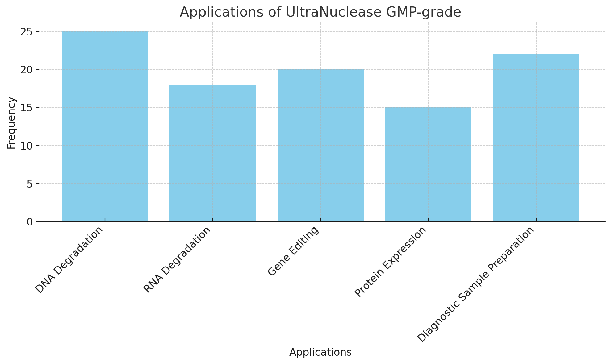
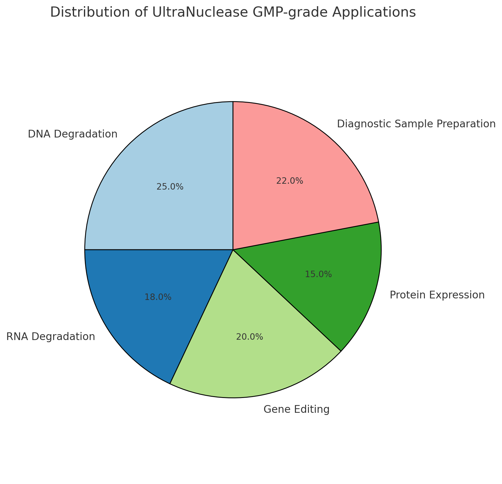
In molecular biology, UltraNuclease GMP-grade is commonly used in various laboratory protocols. Below are some of the key applications:

1. DNA and RNA Degradation

UltraNuclease GMP-grade is frequently used in RNA sequencing and gene expression studies, where it ensures that RNA contamination does not interfere with the results. During sequencing, it is crucial to eliminate DNA or other nucleic acids that may introduce errors into the analysis. For a deeper understanding of RNA sequencing, check out this resource from NCBI.

2. Gene Editing Techniques

In gene editing applications like CRISPR-Cas9, UltraNuclease GMP-grade helps to remove residual nucleic acids following gene editing procedures. The enzyme is essential in ensuring that transfected cells are free from contaminating nucleic acids, which could otherwise impact downstream assays. For more on gene editing, refer to the National Institute of Standards and Technology (NIST).

3. Protein Expression Systems

UltraNuclease GMP-grade is crucial in the production of recombinant proteins in mammalian or bacterial expression systems. By degrading nucleic acids in the culture supernatants, it ensures that the final protein product is not contaminated with unwanted nucleic acids, which could interfere with its activity or stability. Learn more about protein expression at PubMed.

4. Sample Preparation for Diagnostics

UltraNuclease GMP-grade is also used in diagnostics, particularly in the preparation of samples for polymerase chain reaction (PCR) and other molecular diagnostic techniques. The enzyme helps remove any residual DNA or RNA that could lead to false positives or interfere with the accuracy of the diagnostic tests.

AffiZYME® UltraNuclease GMP-grade

Regulatory Compliance and Quality Control

UltraNuclease GMP-grade is manufactured under the highest quality standards, ensuring that it meets all regulatory requirements. These include compliance with GMP guidelines, which are designed to ensure that products are consistently produced and controlled according to quality standards. Quality control tests performed during manufacturing include:

  • Purity Tests: To ensure the absence of contaminants such as residual proteins or endotoxins, which could affect experimental results or product safety.

  • Activity Assays: To measure the enzyme’s ability to degrade nucleic acids efficiently.

  • Sterility Testing: To ensure that the enzyme is free from any microbial contamination, making it safe for use in clinical and research settings.

The rigorous quality control process guarantees that UltraNuclease GMP-grade is both effective and safe for use in clinical research and other highly regulated applications. The FDA’s website offers more information on regulatory processes for enzyme products.

Advantages of UltraNuclease GMP-grade

There are several significant advantages to using UltraNuclease GMP-grade in molecular biology and biotechnology:

  1. High Purity and Specificity: UltraNuclease GMP-grade ensures the efficient degradation of DNA and RNA, ensuring that unwanted nucleic acids are removed while leaving proteins and other critical components intact.

  2. Regulatory Compliance: Produced under strict GMP conditions, UltraNuclease GMP-grade is suitable for use in clinical trials, biopharmaceutical production, and other regulated environments.

  3. Versatility: It can be used in a wide range of applications, from research and diagnostics to large-scale pharmaceutical manufacturing.

  4. Safety: UltraNuclease GMP-grade is thoroughly tested for sterility and endotoxin levels, making it safe for use in clinical and pharmaceutical environments.

For additional insights on the role of enzymes in clinical and pharmaceutical research, the NIH provides a wealth of information.

UltraNuclease GMP-grade in Pharmaceutical Manufacturing

In the pharmaceutical industry, UltraNuclease GMP-grade plays a crucial role in the production of biologic drugs, where purity and contamination-free products are critical. Whether for monoclonal antibody production or gene therapy, ensuring the elimination of nucleic acids is essential for maintaining product quality and safety. Enzyme products like UltraNuclease GMP-grade are vital in the upstream and downstream processing of biologic drugs, ensuring the final product is free from any unwanted nucleic acid material.

UltraNuclease GMP-grade also plays an essential role in the purification process, where its ability to degrade nucleic acids prevents them from interfering with the production of the desired therapeutic proteins.

Research and Future Directions

As research in biotechnology and molecular biology advances, the demand for high-quality enzymes like UltraNuclease GMP-grade is expected to grow. Enzymes are playing an increasingly important role in the advancement of genomics and other high-throughput technologies. By offering greater specificity, stability, and compliance with regulatory standards, UltraNuclease GMP-grade is poised to be a vital tool for researchers and pharmaceutical companies alike.

As the field continues to grow, it is crucial to monitor new advancements in enzyme technologies, ensuring that new innovations align with the latest regulatory guidelines. For more on enzyme research and development, visit the National Science Foundation (NSF).

Conclusion

UltraNuclease GMP-grade is a powerful enzyme that plays a crucial role in biotechnology and pharmaceutical applications. It provides researchers and manufacturers with a reliable tool for nucleic acid degradation, ensuring cleaner, more accurate results in a wide variety of applications. From molecular biology research to the production of biologics, UltraNuclease GMP-grade is an essential tool in modern biotechnology. By adhering to GMP standards, it ensures that it meets the highest regulatory requirements for clinical and therapeutic applications.

For further resources on enzyme technologies, visit PubMed, NCBI, and the World Health Organization (WHO).

Optimizing Immunodetection: The Role of Goat anti-mClover Polyclonal IgG Antibody in Fluorescence-Based Assays

Fluorescence-based assays have become fundamental in many biological and biochemical analyses due to their sensitivity, specificity, and ability to provide quantitative data. These assays depend heavily on the interaction between primary and secondary antibodies to detect antigens, with secondary antibodies like Goat anti-mClover Polyclonal IgG Antibody playing a key role in signal amplification and visualization. This article explores the role of Goat anti-mClover Polyclonal IgG in fluorescence-based assays, with an emphasis on optimizing protocols to ensure robust and reliable results. Additionally, we will highlight applications, technical considerations, and best practices for the optimal use of this antibody.

1. Understanding Fluorescence-Based Immunodetection Assays

Fluorescence-based immunodetection assays are widely used in research and diagnostics to detect specific biomolecules through the use of labeled antibodies. The primary antibody binds to the target antigen, while the secondary antibody, conjugated to a fluorophore, binds to the primary antibody. Upon excitation with a specific wavelength of light, the fluorophore emits light at a longer wavelength, which is captured to visualize the target. This method is employed in a variety of assays, including immunohistochemistry, immunofluorescence, and Western blotting, to detect proteins or other molecules of interest.

The advantages of fluorescence-based assays over traditional methods include their high sensitivity, ability to multiplex, and non-invasive nature, making them ideal for live-cell imaging and dynamic studies of protein-protein interactions. For a more comprehensive understanding of the principles behind these techniques, visit the National Institute of General Medical Sciences.

2. The Goat anti-mClover Polyclonal IgG Antibody

The Goat anti-mClover Polyclonal IgG Antibody is a highly specific secondary antibody designed to recognize and bind to mClover, a green fluorescent protein variant commonly used in recombinant protein tagging. This polyclonal antibody is raised in goats and offers high-affinity binding due to its recognition of the mClover protein’s unique epitopes. The antibody provides an effective means of amplifying the fluorescent signal in assays, making it an essential tool in fluorescence microscopy and other related applications.

The polyclonal nature of the Goat anti-mClover antibody means that it can recognize and bind multiple epitopes of mClover, resulting in stronger signal generation and improved sensitivity. This characteristic is particularly useful in experiments requiring robust signal amplification, such as single-molecule detection and low-abundance target studies. To learn more about the mechanisms of polyclonal antibody production, visit NIH’s Antibody Resources.

3. Fluorescence Labeling and Detection

Fluorescence-based assays rely on the conjugation of secondary antibodies with fluorophores that emit light upon excitation. The Goat anti-mClover Polyclonal IgG Antibody is often conjugated with fluorophores like Alexa Fluor 488, which emit a green fluorescence that matches the emission spectrum of mClover. Choosing the right fluorophore is critical for maximizing signal intensity and ensuring that the emission spectrum matches the detector’s capabilities.

Fluorescence labeling is most commonly used in confocal and wide-field microscopy, flow cytometry, and microplate readers. For a detailed guide to fluorophore selection in immunodetection, refer to Thermo Fisher’s fluorophore selection guide.

4. Optimizing Goat anti-mClover Polyclonal IgG Antibody in Assays

Optimization of the Goat anti-mClover Polyclonal IgG Antibody is essential for obtaining reproducible and reliable results in fluorescence assays. Here, we outline the key parameters that need to be fine-tuned for the best performance.

4.1. Antibody Concentration

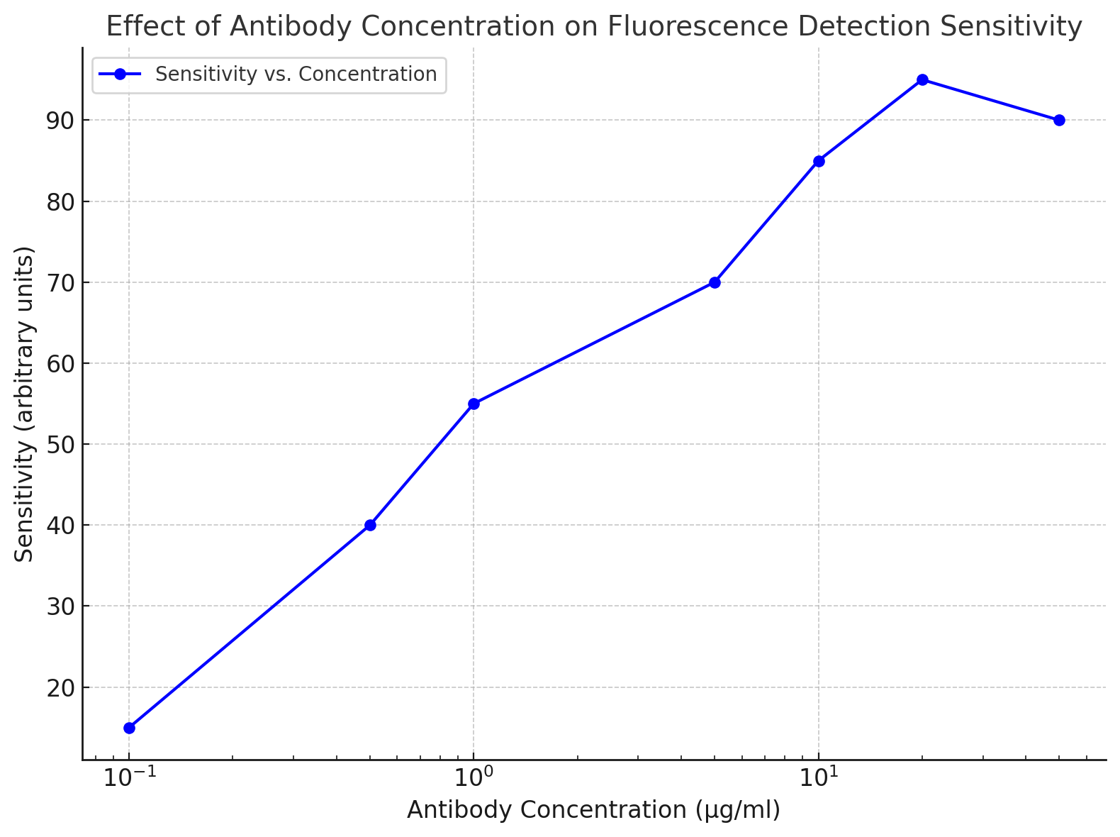
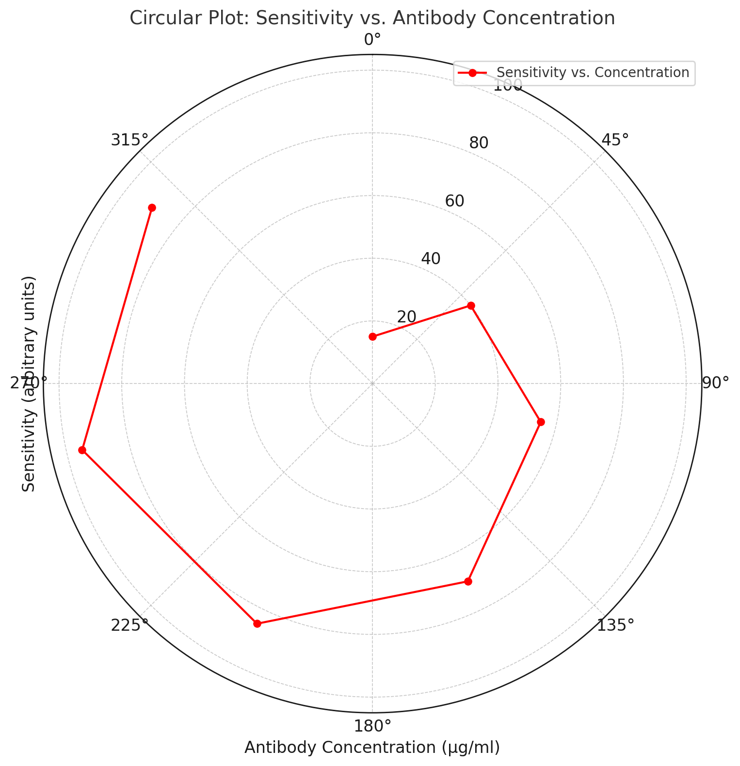
The optimal concentration of the Goat anti-mClover antibody depends on several factors, including the specific assay, target abundance, and fluorophore conjugate. Over-concentration of antibodies can lead to high background fluorescence, while under-concentration may result in weak signals. Titrating the antibody concentration is the most effective way to identify the best working concentration. For titration protocols, refer to ASBMB’s antibody protocols.

AffiAB® Goat anti-mClover  Polyclonal IgG Antibody
4.2. Blocking Agents

Blocking agents are necessary to prevent non-specific binding of antibodies to other components of the sample. Common blocking agents include BSA (Bovine Serum Albumin), Casein, and Tween-20. These agents effectively block sites that may otherwise cause background fluorescence, enhancing the specificity of the assay.

4.3. Incubation Time

Incubation time for the Goat anti-mClover Polyclonal IgG Antibody must be carefully optimized. Too short of an incubation may result in insufficient binding, while overly long incubation can cause excessive background. The optimal incubation time typically ranges from 30 minutes to 1 hour at room temperature, depending on the experimental conditions. For detailed protocol optimizations, consult the University of California’s Immunodetection Protocols.

4.4. Washing Conditions

After antibody incubation, extensive washing is crucial to remove unbound antibodies and reduce non-specific fluorescence. Using buffers such as PBS or TBS helps remove excess antibodies. Gentle washing, typically with 3–4 washes for 5–10 minutes, is recommended to minimize loss of signal. For further washing techniques, see PubMed’s best practices for washing steps.

4.5. Sample Preparation

The integrity of the sample plays a critical role in assay performance. For cell-based assays, cells must be properly fixed and permeabilized to ensure the antibody penetrates and binds the target. For tissue samples, proper fixation and sectioning are crucial for maintaining target integrity. For more information on tissue sample preparation, refer to NIH’s tissue handling guide.

5. Common Applications of Goat anti-mClover Polyclonal IgG Antibody

The Goat anti-mClover Polyclonal IgG Antibody has broad applications in various research fields, particularly in molecular and cell biology.

5.1. Cell Tracking and Migration Studies

In live-cell imaging, mClover is often used to tag proteins of interest. The Goat anti-mClover antibody enables the visualization of these proteins in real time, making it a powerful tool for studying cell migration and dynamic protein localization. This application is critical in cancer research, cell motility studies, and neurobiology.

5.2. Protein Localization and Expression Studies

By tagging proteins with mClover, researchers can visualize the spatial distribution of proteins within cells and tissues. This method is crucial for understanding protein trafficking, organelle function, and cellular responses to external stimuli. For a detailed study of protein localization, check out Cellular Signaling Pathways.

5.3. Transgenic Animal Models

Transgenic animals expressing mClover provide an invaluable model for studying gene expression and protein function in vivo. These models are frequently used in gene therapy, developmental biology, and disease modeling.

6. Future Directions in Fluorescence-Based Immunodetection

The use of the Goat anti-mClover Polyclonal IgG Antibody is continuously evolving with advances in technology. New developments in multi-plexing, where multiple targets are detected simultaneously, offer new insights into cellular processes. The integration of super-resolution microscopy and other advanced imaging techniques further enhances the capabilities of fluorescence-based assays.

Conclusion

The Goat anti-mClover Polyclonal IgG Antibody is a powerful tool for enhancing the sensitivity and specificity of fluorescence-based immunodetection assays. By optimizing factors such as antibody concentration, blocking agents, and incubation times, researchers can significantly improve assay performance. With its wide range of applications in cell biology, protein research, and animal models, this antibody is an essential tool in modern biomedical research. As technology advances, fluorescence-based assays will continue to provide groundbreaking insights into cellular and molecular processes.

The Role of Veterinary Rapid Tests in Zoonotic Disease Surveillance: Detecting Pathogens Before They Spread

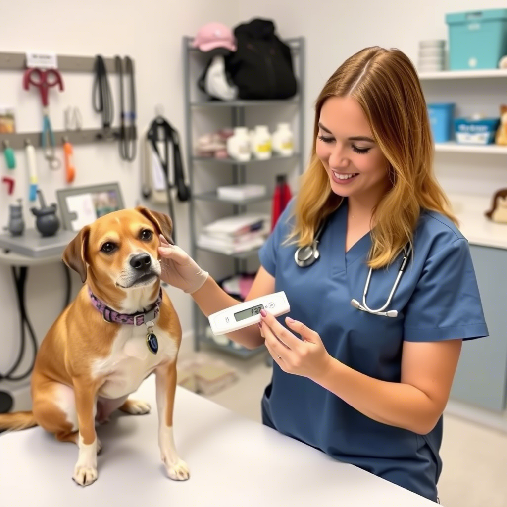
Zoonotic diseases represent a significant threat to global public health, agriculture, and wildlife conservation. These diseases, caused by pathogens that can be transmitted between animals and humans, are responsible for over 60% of emerging infectious diseases, as highlighted by the Centers for Disease Control and Prevention (CDC). Understanding and controlling zoonotic diseases are essential for preventing potential outbreaks that could lead to widespread health crises. One of the most vital tools in combating these diseases is the use of veterinary rapid tests for pathogen detection. These diagnostic technologies allow for early identification of pathogens in animals, reducing the risk of transmission to humans and other animals.

In this article, we explore the importance of veterinary rapid tests in zoonotic disease surveillance, detailing how they are used to detect pathogens before they can spread and the critical role they play in public health and animal health management.

What Are Zoonotic Diseases?

Zoonotic diseases are diseases that can be transmitted between animals and humans, either directly or indirectly. Some zoonotic diseases, such as Avian Influenza, Rabies, Brucellosis, and Hantavirus, are well-known for their capacity to jump from animals to humans, causing significant health problems and sometimes leading to pandemics. According to the World Health Organization (WHO), zoonotic diseases are responsible for approximately 75% of emerging infectious diseases in humans.

Transmission pathways of zoonotic diseases can vary, including direct contact with infected animals, consumption of contaminated animal products, and insect vectors that carry pathogens between species. Surveillance and early detection of zoonotic pathogens are critical in preventing such diseases from spreading across populations and geographical regions.

The Importance of Early Detection in Zoonotic Disease Control

Detecting zoonotic pathogens early is crucial in preventing the spread of disease and reducing the impact on both animal and human populations. Veterinary rapid tests are a key component of disease surveillance programs, providing quick and accurate results that inform immediate intervention strategies. Rapid detection allows veterinarians to isolate infected animals, implement control measures, and prevent further transmission.

Early detection can also help identify the reservoirs of zoonotic diseases, which are often wildlife species or domestic animals. By understanding where pathogens are originating, health officials can implement measures to limit human exposure and reduce the risk of zoonotic diseases becoming endemic in human populations.

For example, rabies is a fatal viral disease that can be rapidly detected in animals through specialized tests. Timely diagnosis helps to prevent human exposure and allows for the vaccination of at-risk animal populations. Similarly, the detection of Avian Influenza in poultry can prevent human cases by allowing authorities to cull infected animals and restrict trade and movement.

AffiVET® Canine Brucella Antibody Rapid Test Kit

Types of Veterinary Rapid Tests for Zoonotic Disease Detection

Several types of veterinary rapid tests are used to detect zoonotic pathogens in animals, and each plays a unique role in disease surveillance. These diagnostic tests are typically categorized into three major groups:

  1. Antigen Detection Tests

    • These tests detect specific proteins (antigens) on the surface of pathogens. For instance, Avian Influenza virus can be detected using antigen-based rapid tests, helping to prevent the virus from spreading to humans and other animals. Antigen tests are widely used because they are relatively inexpensive and easy to use in the field. The Centers for Disease Control and Prevention (CDC) provides guidelines for the proper use of these diagnostic tools in controlling animal outbreaks.

  2. Polymerase Chain Reaction (PCR) Tests

    • PCR testing is a molecular technique used to amplify and detect the genetic material of pathogens. PCR tests are highly sensitive and specific, making them ideal for detecting pathogens like Brucella, FMDV (Foot-and-Mouth Disease Virus), and Ebola. PCR allows for the detection of even low quantities of the pathogen’s DNA or RNA, which is especially useful in early-stage infections where pathogen loads may be low. The National Institutes of Health (NIH) has developed various protocols for PCR testing in veterinary diagnostics, which are used by veterinarians worldwide.

  3. Immunoassays

    • Immunoassays are tests that use antibodies to detect pathogens or their components. These tests are commonly used for detecting Bovine Tuberculosis (TB), Leptospirosis, and Lyme disease in animals. Immunoassays provide quick results, and some tests can be performed on-site without the need for specialized laboratory equipment. The USDA National Animal Health Monitoring System (NAHMS) has evaluated immunoassays’ effectiveness in diagnosing bovine diseases and has supported their use for large-scale surveillance in cattle populations.

Each of these tests offers distinct advantages in terms of speed, accuracy, and cost-effectiveness. They play a central role in identifying infected animals and preventing the further spread of zoonotic diseases to humans, livestock, and wildlife.

The Role of Rapid Tests in Global Disease Surveillance

Veterinary rapid tests are integral components of global disease surveillance systems. These systems help to monitor the spread of zoonotic diseases across geographic regions and ensure timely responses when outbreaks occur. Surveillance programs, such as those run by the World Organization for Animal Health (OIE) and CDC, track the incidence of zoonotic diseases in animal populations and provide critical data to inform public health interventions.

The integration of rapid testing into global surveillance frameworks, such as the One Health Initiative, ensures that zoonotic diseases are monitored from animal populations to humans. The One Health approach acknowledges that the health of humans, animals, and the environment are interconnected, and it emphasizes the need for collaboration between veterinary, medical, and environmental health sectors to effectively address zoonotic threats. The CDC’s One Health Office coordinates efforts to prevent and control zoonotic diseases globally.

Rapid Tests in Disease Control and Containment

Once a pathogen is detected using veterinary rapid tests, immediate containment measures can be implemented to limit the spread of the disease. These measures can include the quarantine of infected animals, culling of affected livestock, and restriction of trade to prevent the movement of infected animals and animal products.

For example, the rapid detection of Brucellosis in cattle can lead to the immediate isolation of infected herds and the testing of surrounding animals to prevent further transmission. Similarly, Rabies detection allows for targeted vaccination campaigns and public awareness efforts to reduce the risk of human exposure.

The Food and Agriculture Organization (FAO) works in partnership with national governments to support the implementation of disease control measures in response to positive rapid test results. The FAO’s Emergency Prevention System for transboundary animal diseases (EMPRES) focuses on early warning systems to identify outbreaks of zoonotic diseases and help deploy rapid response teams.

Challenges and Limitations of Veterinary Rapid Tests

While veterinary rapid tests have proven to be invaluable in detecting zoonotic pathogens, there are several challenges associated with their use. One significant limitation is the sensitivity of some tests, particularly in cases where pathogens exhibit genetic diversity or when the pathogen load in an animal is low. For example, African Swine Fever (ASF) virus can be difficult to detect early in the course of infection, as the virus’s genetic material can vary between strains.

Additionally, logistical challenges such as limited access to testing resources in rural or remote areas can hinder the widespread adoption of these technologies. Access to trained personnel, cold storage for reagents, and reliable transport networks for samples are essential for ensuring that rapid tests can be effectively deployed in the field.

The National Science Foundation (NSF) has funded research into new technologies that aim to address some of these limitations, including CRISPR-based diagnostics and nanotechnology-enabled rapid tests. These advancements hold promise for improving the accuracy, sensitivity, and accessibility of veterinary rapid tests.

Future Directions in Veterinary Rapid Testing

As new zoonotic diseases continue to emerge, the need for advanced diagnostic tools will only increase. Researchers are exploring innovative solutions to enhance the speed, accuracy, and cost-effectiveness of veterinary rapid tests. The integration of digital technologies, such as smartphone-based diagnostic platforms, is also expanding the possibilities for field diagnostics in remote regions.

The National Institute of Standards and Technology (NIST) is working on improving the performance of diagnostic tests, ensuring they meet global standards for reliability and reproducibility. The future of veterinary rapid tests lies in a multi-disease testing approach, which could detect a broad range of pathogens simultaneously, making surveillance more efficient and comprehensive.

Conclusion

Veterinary rapid tests are essential tools in the surveillance and control of zoonotic diseases. By enabling early detection of pathogens, these tests help prevent outbreaks and mitigate their impact on human and animal populations. The continued development of these diagnostic tools, coupled with global efforts in disease surveillance, will enhance our ability to respond to emerging zoonotic threats. Through effective early detection, we can protect public health, safeguard wildlife, and reduce the economic burden of zoonotic diseases worldwide.


Sources for Further Reading:

Advancements in Bead-Based Assays for Multiplexed Diagnostic Testing: How Simultaneous Detection Improves Diagnostic Accuracy

Introduction

Bead-based assays represent a groundbreaking advancement in diagnostic testing. These assays enable the detection of multiple analytes simultaneously in a single sample, offering enhanced diagnostic accuracy and efficiency. By utilizing microbeads coated with specific capture agents, bead-based assays provide significant benefits over traditional assays, including higher sensitivity, reduced sample volume requirements, and faster turnaround times. These advancements are revolutionizing the way we approach disease detection and monitoring, particularly in clinical diagnostics, environmental testing, and disease surveillance.

The key feature of bead-based assays is their multiplexing capability—the ability to test for multiple biomarkers in a single test. This not only increases the throughput of diagnostic systems but also enables more comprehensive analyses in clinical practice. The simultaneous detection of multiple biomarkers can lead to earlier and more accurate diagnosis, better patient outcomes, and more effective disease monitoring.

Bead-Based Assays: A Technological Overview

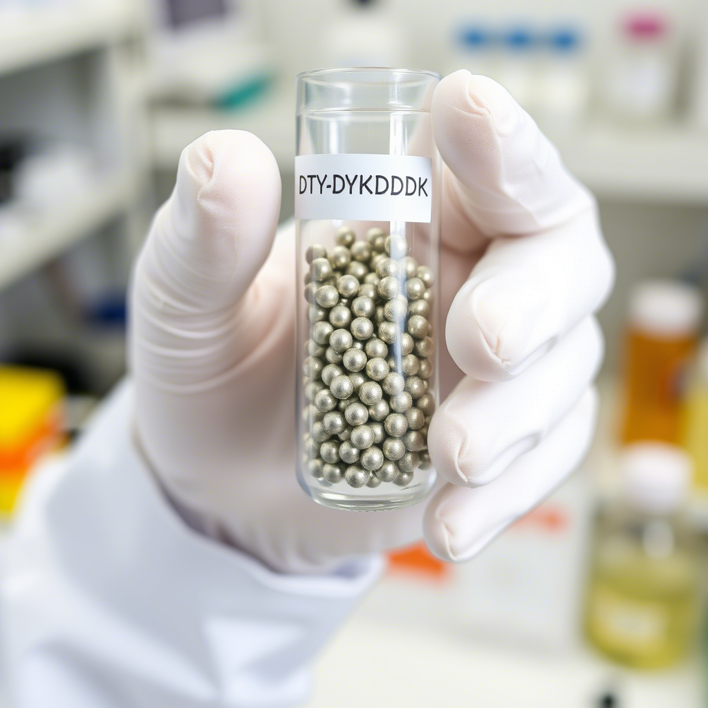
Bead-based assays are built around microbeads that are functionalized with various ligands, such as antibodies, aptamers, or peptides, to specifically bind to target analytes. The beads serve as solid-phase supports for capturing the biomolecules of interest. After exposure to a sample, the beads bind to their specific targets, and the presence of these analytes is detected using a range of optical or electrochemical detection methods. These assays can be performed in a multiplex format, meaning that different beads, each conjugated with a specific ligand, can be used in parallel to detect multiple targets at the same time.

The multiplexing capability is achieved by assigning distinct physical properties, such as size or color, to the beads. This allows each bead to carry a unique identification tag that can be used to differentiate between multiple analytes in a single sample. Detection systems such as flow cytometry, fluorescence-based techniques, and colorimetric detection are commonly used to analyze the beads. One key advantage of bead-based assays is their ability to detect analytes in complex samples with minimal interference, providing reliable results in clinical diagnostics, environmental monitoring, and other applications.

Key Advancements in Bead-Based Assays

Over the past decade, several technological advancements have significantly enhanced the capabilities of bead-based assays. These innovations include improvements in assay sensitivity, the incorporation of new detection technologies, the integration of microfluidics, and the automation of multiplex assays. Below are some of the notable advancements in this area:

1. Enhanced Sensitivity Through Advanced Surface Chemistry

Advancements in surface chemistry have been a game-changer for bead-based assays, allowing for more efficient binding of capture agents to the beads and providing enhanced specificity for target biomarkers. Researchers have developed novel surface modifications and functionalization techniques that increase the binding capacity of the beads, ensuring that even low-abundance analytes can be effectively captured and detected.

For example, researchers at University of California, Berkeley have demonstrated how the use of high-density capture agents improves the detection sensitivity of bead-based assays for low-abundance biomarkers, enabling the detection of rare pathogens or early-stage disease markers. This advancement is particularly useful in fields such as cancer biomarker detection and infectious disease diagnostics.

2. Integration of Nanotechnology for Increased Multiplexing

The incorporation of nanotechnology into bead-based assays has significantly increased their multiplexing capabilities. By integrating nanoparticles with different optical properties, it is now possible to detect and analyze dozens of biomarkers simultaneously in a single test. Nanoparticles, such as gold nanoparticles and quantum dots, can be engineered to emit specific wavelengths of light or exhibit unique color properties, allowing for the simultaneous detection of a wide array of targets.

Studies conducted by researchers at MIT have demonstrated the use of quantum dots in bead-based assays, enabling the detection of over 100 different biomarkers in a single sample. This level of multiplexing is crucial in the development of diagnostic tests for complex diseases like cancer, where multiple biomarkers need to be measured simultaneously to provide a comprehensive analysis.

3. High-Throughput and Automation

Automation is one of the most important advancements in bead-based assays, enabling high-throughput testing in clinical and research settings. Bead-based assays are increasingly being integrated into automated platforms that allow for the processing of hundreds or even thousands of samples per day. These platforms can automatically dispense beads, incubate samples, wash the beads, and read the detection signals, reducing the need for manual intervention and minimizing the risk of human error.

The FDA has approved several automated bead-based multiplexed diagnostic systems for use in clinical diagnostics, including platforms for detecting infectious diseases, cancer biomarkers, and autoimmune diseases. Automation not only improves throughput but also increases consistency and reproducibility across different laboratories and settings.

4. Microfluidic Integration for Efficient Sample Processing

Microfluidics, the science of manipulating small volumes of fluids, has been integrated with bead-based assays to increase their efficiency and decrease the cost of diagnostic tests. Microfluidic devices allow for the precise control and manipulation of fluids on a microscopic scale, enabling faster sample processing and reducing the amount of reagent and sample required for testing.

Researchers at Stanford University have developed microfluidic systems that integrate bead-based assays for rapid point-of-care diagnostics. These systems can perform multiple tests on a single sample within minutes, making them ideal for use in resource-limited settings or for on-site diagnostics in remote areas.

Applications of Bead-Based Assays

Bead-based assays have a wide range of applications, particularly in the fields of clinical diagnostics, environmental monitoring, and disease surveillance. Their ability to detect multiple biomarkers simultaneously makes them especially useful for complex diagnostic scenarios where a single test may not provide enough information.

1. Infectious Disease Detection

In the field of infectious diseases, bead-based assays are invaluable for the rapid detection of pathogens. Multiplexed bead assays can test for multiple pathogens in a single sample, reducing the need for multiple diagnostic tests and enabling faster decision-making in clinical settings.

The CDC has implemented bead-based assays to improve diagnostic accuracy in detecting viral and bacterial infections, including HIV, Zika virus, and respiratory pathogens like influenza and COVID-19. These assays offer a rapid and accurate means of diagnosing infections, allowing healthcare providers to initiate treatment more quickly.

AffiBEADS® Anti-DYKDDDDK (Flag) Magnetic Beads

2. Cancer Diagnosis and Monitoring

Bead-based assays have become an essential tool for cancer biomarker detection. By measuring a panel of cancer-specific biomarkers in a single sample, these assays provide a more comprehensive picture of a patient’s condition. This is especially important in cancers that involve multiple pathways, such as lung, breast, and colorectal cancer.

MD Anderson Cancer Center has employed bead-based assays to monitor the progression of cancer and detect tumor markers in blood samples. This approach allows for earlier detection of recurrence and better tracking of disease progression, leading to more personalized treatment strategies.

3. Autoimmune Diseases

In autoimmune diseases, bead-based assays are used to detect a broad range of autoantibodies that are indicative of conditions like lupus, rheumatoid arthritis, and multiple sclerosis. By simultaneously detecting multiple autoantibodies in a single test, bead-based assays provide a more accurate and comprehensive diagnosis, reducing the time and cost associated with traditional testing methods.

The National Institute of Arthritis and Musculoskeletal and Skin Diseases (NIAMS) has supported the use of bead-based assays in autoimmune disease research, highlighting their ability to detect a wide array of autoantibodies that are critical for understanding disease progression and treatment response.

4. Environmental Monitoring and Food Safety

Beyond clinical diagnostics, bead-based assays are increasingly used in environmental monitoring and food safety testing. These assays can detect contaminants, such as pathogens, toxins, and heavy metals, in water, soil, and food products. Their ability to perform multiplexed detection makes them particularly suitable for environmental surveillance, where multiple contaminants may be present at low levels.

The EPA has been involved in developing bead-based assays for the detection of harmful microorganisms in water sources, providing faster results than traditional methods and enabling more effective public health protection.

Challenges and Future Directions

While bead-based assays offer many advantages, several challenges remain. One of the primary challenges is the complexity of assay development, particularly when working with large numbers of biomarkers. The specificity of the capture agents must be carefully optimized to avoid cross-reactivity, and the detection systems must be highly sensitive to ensure accurate results.

Moreover, as the number of biomarkers included in a multiplexed assay increases, the complexity of data interpretation also grows. Advanced bioinformatics tools and machine learning algorithms are being developed to assist with the analysis of large datasets generated by multiplexed assays.

The future of bead-based assays looks promising, with ongoing research into new materials, advanced detection technologies, and portable devices. With the continued advancements in nanotechnology and microfluidics, it is likely that bead-based assays will become even more efficient and widely used in both clinical and environmental applications.

Conclusion

Bead-based assays for multiplexed diagnostic testing represent a major advancement in the field of diagnostics, offering improved sensitivity, higher throughput, and the ability to detect multiple biomarkers in a single test. These assays are revolutionizing the way we diagnose and monitor a wide range of diseases, from infectious diseases to cancer and autoimmune disorders. As technology continues to evolve, bead-based assays will play an increasingly important role in clinical diagnostics, environmental monitoring, and public health surveillance.

For further information on the latest developments in bead-based assay technologies, visit trusted resources from NIH, Johns Hopkins University, and Stanford University.

Newcastle Disease Virus (NDV): Moleculaire Inzichten, Surveillance en Beheersing in Vogelpopulaties

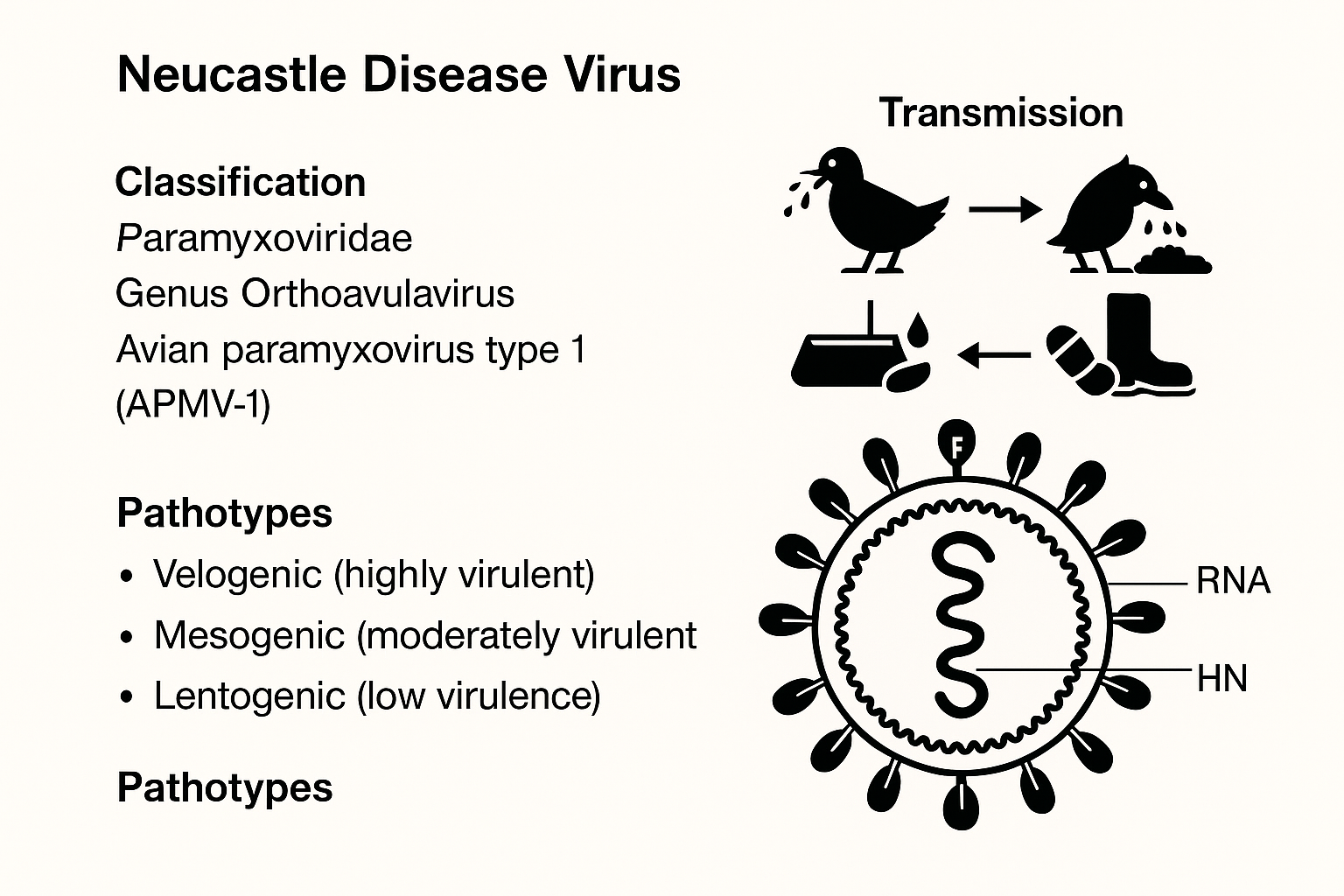
Newcastle Disease Virus (NDV) is een wereldwijd verspreid aviër paramyxovirus dat ernstige uitbraken veroorzaakt in vogelpopulaties. Dit RNA-virus vormt een grote uitdaging voor pluimveebedrijven, volières en broederijen, vooral in gebieden waar bioveiligheid en vaccinatieprogramma’s inconsistent zijn. De infectie wordt veroorzaakt door stammen van Avian Paramyxovirus serotype 1 (APMV-1) en varieert in ernst, afhankelijk van het genotype, gastheersoort en immuunstatus van de vogels.

Virale Classificatie en Genomica

NDV behoort tot het geslacht Orthoavulavirus, familie Paramyxoviridae, en orde Mononegavirales. Het volledige genoom van NDV varieert van 15.186 tot 15.198 nucleotiden, afhankelijk van de stam (NCBI Genome). Het genoom codeert zes structurele eiwitten:

  • NP (nucleocapside)

  • P (fosfoproteïne)

  • M (matrixeiwit)

  • F (fusie-eiwit)

  • HN (hemagglutinine-neuraminidase)

  • L (grote polymerase)

De F-eiwit-klievingsplaats is een belangrijke determinant van virulentie. Virulente stammen hebben meerdere basische aminozuren op deze plaats, waardoor klieving door host-proteasen mogelijk wordt, wat leidt tot systemische verspreiding van het virus (Cornell University Avian Diagnostic Lab).

Pathotypen van NDV

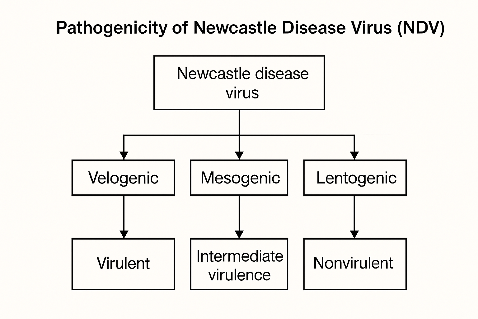
NDV-stammen worden ingedeeld in drie hoofdpathotypen op basis van klinische symptomen en sterfte bij kippen:

  • Velogeen: Zeer virulent, veroorzaakt neurologische, respiratoire en gastro-intestinale symptomen. Vaak snelle sterfte.

  • Mesogeen: Matige virulentie, gematigde ademhalings- en zenuwverschijnselen.

  • Lentogeen: Lage virulentie; meestal gebruikt in levend verzwakte vaccins (Merck Veterinary Manual).

Deze classificaties worden gevalideerd via biologische tests zoals de intracerebrale pathogeniciteitsindex (ICPI) en de gemiddelde sterftijd (MDT) in embryonale eieren (FAO ND Manual).

Natuurlijk Gastheerbereik en Virale Uitscheiding

NDV infecteert meer dan 250 vogelsoorten, zowel gedomesticeerde als wilde. Kippen zijn het meest vatbaar, maar het virus treft ook kalkoenen, duiven, fazanten, eenden, ganzen en wilde vogels zoals aalscholvers en sterns. Geïnfecteerde vogels scheiden het virus uit via:

  • Uitwerpselen

  • Neusafscheidingen

  • Veerdander

  • Besmette oppervlakken (voeders, waterleidingen, kooien)

Virale uitscheiding begint ongeveer 2–5 dagen na infectie en kan weken aanhouden, afhankelijk van de soort en stam (USGS National Wildlife Health Center).

Transmissiemechanismen

NDV verspreidt zich via:

  • Direct contact tussen vogels

  • Fomieten (schoenen, voertuigen, gereedschap, voer)

  • Aerosoloverdracht

  • Verticale transmissie (zeldzaam en slecht gedocumenteerd)

Verspreiding over lange afstand vindt ook plaats via trekvogels en slecht gereinigde transportcontainers voor levend pluimvee. Het virus is milieustabiel, vooral in organisch materiaal en bij lage temperaturen (USDA Biosecurity Guide).

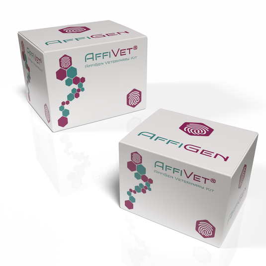
AffiVET® Newcastle Disease Virus (NDV) Antigen Rapid Test

Moleculaire Detectie en Surveillance

Nauwkeurige detectie van NDV is essentieel voor controle en beheersing. Veelgebruikte methoden zijn:

  • Real-time RT-PCR: Gericht op het fusiegen voor snelle identificatie (CDC Protocols)

  • Virusisolatie: In SPF-eieren

  • Hemagglutinatieremming (HI)-test

  • Next-generation sequencing: Voor genomische monitoring en variantenanalyse (NCBI Virus).

Surveillance wordt ondersteund door netwerken zoals National Animal Health Laboratory Network (NAHLN) en National Poultry Improvement Plan (NPIP).

Vaccinontwikkeling en Toepassing

Vaccinatie blijft de meest effectieve methode om NDV-uitbraken te voorkomen. Huidige vaccins omvatten:

  • Levend verzwakte vaccins (LaSota, B1)

  • Geïnactiveerde olie-emulsievaccins

  • Recombinante vectorvaccins (bijv. fowlpox-NDV)

Toediening gebeurt via:

  • Aerosolverneveling

  • Drinkwater

  • Oog- of neusdruppels

Onderzoek richt zich ook op subunitvaccins, DNA-gebaseerde constructen en plantexpressie van antigenen voor schaalbare en goedkope productie (ARS USDA Vaccine Research).

Omgevingsstrategieën en Management

NDV-uitbraken kunnen hele vogelpopulaties verwoesten als bioveiligheidsprotocollen niet worden gevolgd. Belangrijke maatregelen:

  • Isolatie van nieuwe koppels

  • Quarantaine van zieke of blootgestelde vogels

  • Beperkte toegang tot stallen

  • Desinfectie van materiaal en infrastructuur

  • Gebruik van NDV-vrij voer en strooisel

De campagne Biosecurity for Birds van de USDA biedt gratis materialen en instructies voor pluimveehouders (Biosecurity Portal).

Wereldwijde Epidemiologie en Wilde Vogelmonitoring

NDV is endemisch in delen van Afrika, Azië en Latijns-Amerika. In Noord-Amerika en Europa zijn uitbraken zeldzaam maar mogelijk, vooral via de handel in vogels en contact met wilde soorten. De Wereldorganisatie voor Diergezondheid (WOAH) classificeert het als een ziekte met grensoverschrijdend risico (WOAH Disease Card).

Monitoring van trekvogels gebeurt via netwerken zoals Bird Banding Laboratory (USGS) en USDA Wildlife Services.

Onderzoekstrends en Innovaties

NDV-onderzoek richt zich op:

  • Genomische plasticiteit en recombinatie

  • CRISPR-technologieën voor genbewerking

  • Reverse genetics voor verzwakte vaccins

  • Mucosale immuniteit bij vogels

  • Overlevingsmodellen van het virus in de omgeving

Belangrijke onderzoekscentra:

Conclusie

Newcastle Disease Virus blijft een constante bedreiging voor vogelpopulaties wereldwijd. Door wetenschappelijke samenwerking, genetische monitoring en strategische vaccinatie kunnen verspreiding en economische schade beperkt worden. Kennis van NDV op moleculair, ecologisch en populatieniveau is essentieel voor effectieve preventie op lokaal en internationaal niveau.

Annexine A2 ELISA: Technische Gids voor Eiwitkwantificatie in Biologische Systemen

De Annexine A2 ELISA wordt veel gebruikt in biologisch en biochemisch onderzoek om de concentratie van het Annexine A2 (ANXA2)-eiwit te meten in monsters zoals serum, plasma, celkweek-supernatanten en weefsellysaten. Dit eiwit speelt een rol in verschillende biologische systemen, waaronder membraantransport, cytoskelet-interacties en extracellulaire signalering. De ELISA-methode, gebaseerd op immunologische specificiteit en signaalversterking, maakt nauwkeurige kwantificatie mogelijk in uiteenlopende experimentele opstellingen.

Voor basisinformatie over dit eiwit, zie NCBI Gene – ANXA2, en voor structuurdata RCSB Protein Data Bank.

Structuur en Rol van Annexine A2

Annexine A2 is een calciumafhankelijk fosfolipide-bindend eiwit dat een centrale rol speelt in vesiculair transport en membraan-cytoskelet dynamiek. Het bevat een geconserveerd C-terminaal domein dat typisch is voor de annexinefamilie en een uniek N-terminaal motief dat zorgt voor heterotetramere vorming met S100A10.

Functionele studies op nih.gov wijzen op betrokkenheid bij membraanherstel, endocytische routes en organisatie van membraanreceptoren. Voor meer over annexine-eiwitten, raadpleeg PubMed Central.

Principe van de ELISA-techniek

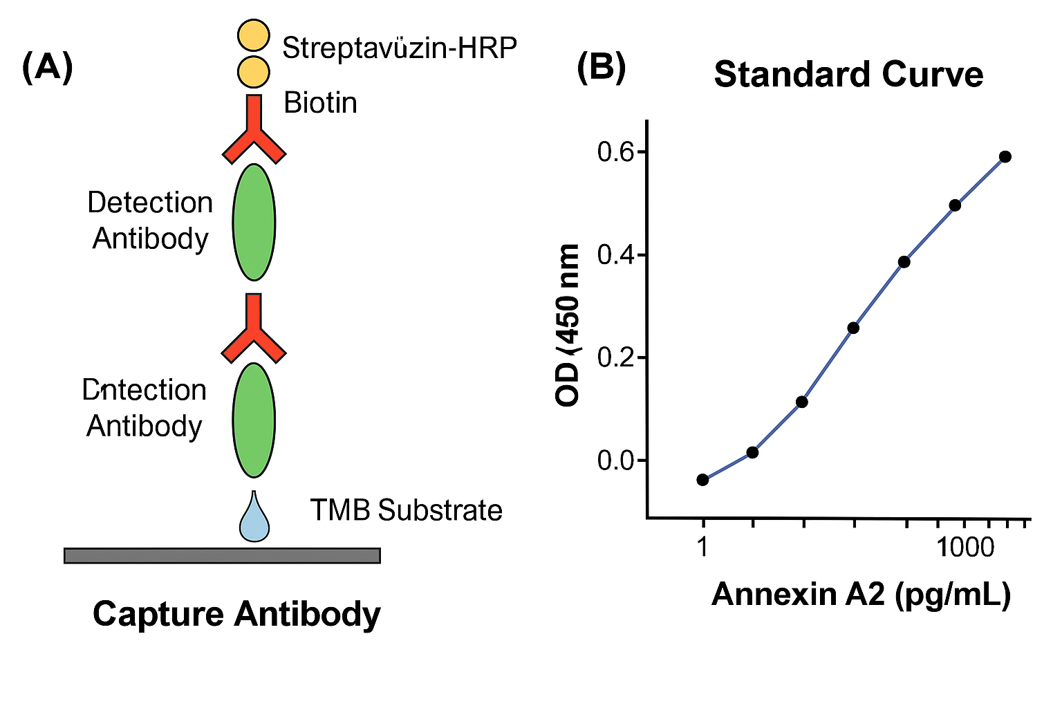
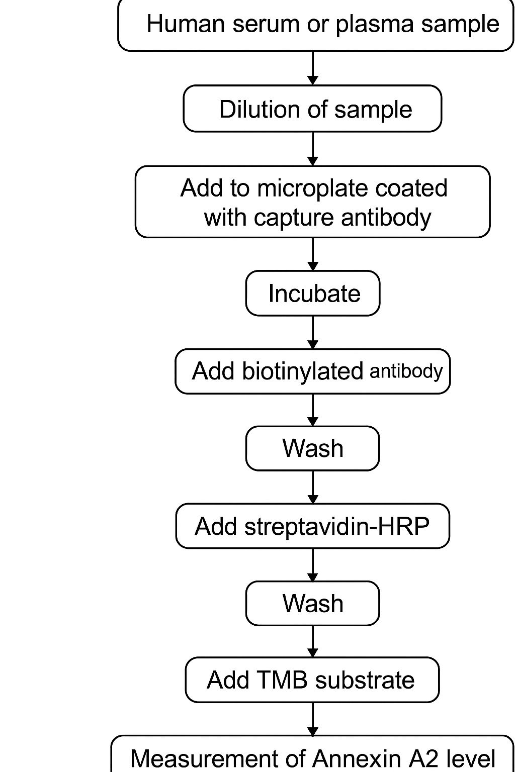
De Annexine A2 ELISA volgt meestal het sandwich ELISA-formaat, bekend om hoge specificiteit en gevoeligheid. Het protocol omvat:

  1. Een 96-wells plaat gecoat met vang-antilichamen tegen Annexine A2

  2. Toevoeging van monsters of standaard-eiwit

  3. Een gebiotinyleerd detectie-antilichaam dat aan een ander epitoop bindt

  4. Enzymgekoppeld streptavidine (meestal HRP)

  5. Een kleurreactie met TMB-substraat

  6. Kwantificatie bij 450 nm met een plaatlezer

Zie Assay Guidance Manual voor details over protocolontwikkeling.

Monstertypes en Voorbewerking

Annexine A2 kan worden gemeten in verschillende biologische matrices:

  • Menselijk of muis serum/plasma

  • Conditioned medium van celkweken

  • Weefselhomogenaten

  • Cellysaten van adherente of suspensiecellijnen

Zie protocollen van laboratoria op jhu.edu, umich.edu en ucsf.edu voor details.

Voor weefsellysaten is het essentieel om lage temperaturen aan te houden en proteaseremmers te gebruiken om eiwitafbraak te voorkomen. Bufferkeuze hangt af van het weefseltype, zoals besproken in ncbi.nlm.nih.gov.

Analyserange en Gevoeligheid

Annexine A2 ELISA-kits detecteren meestal concentraties tussen 10 pg/mL en 2 ng/mL. Hoge gevoeligheid wordt bereikt via:

  • Monoklonale antilichamen met hoge affiniteit

  • Lage achtergrondruis

  • Strikte protocolnauwkeurigheid

Valideringsrichtlijnen zoals LOD, lineair bereik, en herhaalbaarheid zijn beschikbaar op fda.gov en PubMed.

Opslag en Stabiliteit van Reagentia

Annexine A2 ELISA-kits bevatten temperatuurgevoelige reagentia:

  • Bewaar platen en antilichamen bij 2–8°C

  • Vermijd herhaald invriezen en ontdooien

  • Gebruik gereconstitueerde reagentia binnen 24 uur

Zie opslagrichtlijnen van onderzoekscentra op cornell.edu, osu.edu en wisc.edu.

AffiELISA® Mouse Annexin A2 ELISA [ Anxa2]

Toepassingen van Annexine A2-Detectie

1. Membraanorganisatie

Annexine A2 bindt fosfatidylserine op calciumafhankelijke wijze. Het eiwit is actief bij:

  • Exocytose

  • Membraanfusie

  • Endosoomtransport

Zie cellimagelibrary.org en nih.gov voor meer info.

2. Interactie met S100A10

Annexine A2 vormt een heterotetramer met S100A10, wat leidt tot positionering aan het celoppervlak. Deze interactie beïnvloedt:

  • Ionkanaaltransport

  • Cytoskeletorganisatie

  • Exocytisch transport

Zie PMC2932715.

3. Differentiatie en Ontwikkeling

Annexine A2 komt tot expressie tijdens embryonale ontwikkeling, celcommitment en celrijping. Onderzoekslaboratoria bij stanford.edu en duke.edu bestuderen deze processen.

Protocoloptimalisatie

Blokkeringsbuffers

Gebruik BSA, caseïne of normaal serum om niet-specifieke binding te voorkomen. Zie de Assay Guidance Manual.

Incubatietijden

  • Coating: Nacht bij 4°C

  • Monsteraanbinding: 1–2 uur kamertemperatuur

  • Detectie: 1 uur

  • Substraatontwikkeling: 15–30 minuten

Gedetailleerde kinetiek staat op nih.gov.

Data-analyse

Gebruik voor standaardkrommen:

  • Vierparametermodellen (4-PL)

  • Lineaire regressie van log-getransformeerde waarden

Zie analysetools op uchicago.edu, r-project.org, of niddk.nih.gov.

Kruisreactiviteit

Annexine A2 ELISA’s zijn hoog specifiek, met minimale kruisreactiviteit naar Annexine A1 of A5. Validatiedata zijn beschikbaar op antibodyregistry.org en ncbi.nlm.nih.gov.

Veiligheid

Volg standaardpraktijken van biosafety level 2 (BSL-2), vooral bij menselijke monsters. Veiligheidsrichtlijnen zijn beschikbaar via osha.gov en epa.gov.

Problemen en Oplossingen

  • Geen signaal: Controleer of het detectie-antilichaam actief is; controleer TMB-versheid

  • Hoge achtergrond: Gebruik vers blokbuffer en intensievere wasstappen

  • Samengedrukte kromme: Verdun monsters zodat ze binnen het bereik vallen

Zie de ELISA Troubleshooting Manual – NIH.

Slotopmerkingen

De Annexine A2 ELISA is een waardevol instrument voor onderzoekers die eiwitexpressie willen meten in complexe biologische systemen. Het biedt nauwkeurigheid, betrouwbaarheid en reproduceerbare resultaten, met ondersteuning van methodologische richtlijnen van instellingen zoals nih.gov, harvard.edu, en ucsf.edu.