
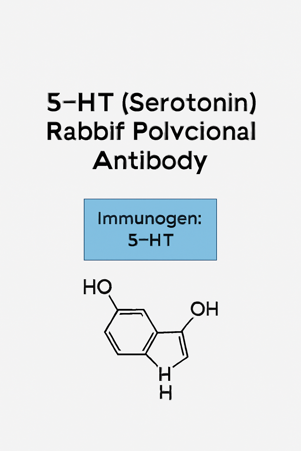
In contemporary neuroscience, gastrointestinal, endocrine and oncological research, the neurotransmitter Serotonin (5-hydroxytryptamine, 5-HT) plays a central role in both physiological signalling and pathology. A robust reagent to detect serotonin content within cells or tissues is the rabbit polyclonal anti-5-HT antibody (often described as “anti-5-HT (serotonin) rabbit polyclonal antibody”) which facilitates immunohistochemistry (IHC), immunofluorescence (IF), immunocytochemistry (ICC) and sometimes quantitative immunoassays.
Below we cover target biology, antibody-selection parameters, protocol considerations, validation and quality control, research applications, troubleshooting, and recommended keyword integration for SEO and indexing.
Target Biology: Serotonin (5-HT) in DetailBiosynthesis, storage, and distribution
Serotonin (5-HT) is synthesised primarily from the amino acid L-tryptophan via the enzymes tryptophan hydroxylase (TPH) and aromatic L-amino acid decarboxylase. Its distribution includes a substantial peripheral pool (notably in enterochromaffin (EC) cells of the gastrointestinal (GI) tract) as well as central nervous system (CNS) serotonergic neurons (for instance the raphe nuclei) and platelet stores. A widely cited review describes how “most serotonin is found outside the CNS” and how nearly all 15 serotonin receptor subtypes have both central and peripheral expression. cdr.lib.unc.edu+2bionumbers.hms.harvard.edu+2
In the GI tract, EC cells are the dominant source of 5-HT; studies estimate ~90–95% of the body’s serotonin is produced in the gut. diposit.ub.edu
Receptor systems & signalling
Serotonin exerts its functions via a diverse family of receptors (5-HT1 through 5-HT7, with ~15 known sub-types) that are primarily G protein-coupled receptors (GPCRs), except for the 5-HT₃ ligand-gated ion channel. wiki.ostrowonline.usc.edu+1
These receptors regulate a vast array of physiological processes: mood regulation, sleep, appetite, gastrointestinal motility, platelet aggregation, vascular tone, and immune cell responses. For example, the “expanded serotonin biology” review outlines regulation of cardiovascular, pulmonary, GI, genitourinary and CNS systems. cdr.lib.unc.edu+1
Pathophysiological relevance
Aberrant serotonergic signalling is implicated in multiple disorders: mood disorders (depression, anxiety), GI disorders (irritable bowel syndrome), neurodegenerative and developmental disorders, carcinoid tumours (serotonin-producing), and emerging evidence in oncology and immunology (for example, serotonin transporter expression modulating tumour immunity). Cell+1
Given the wide physiological relevance and diverse tissue distribution of serotonin, accurate immunodetection of 5-HT is of high research value.
Choosing a Rabbit Polyclonal Anti-5-HT (Serotonin) Antibody
When selecting a rabbit polyclonal antibody against serotonin (i.e., a reagent raised in rabbit, polyclonal IgG fraction recognising serotonin or a serotonin-carrier conjugate), key technical parameters should be evaluated:
Host species & clonality
-
Host: Rabbit. Rabbits are preferred for polyclonal antibodies because they generate high titres and diverse epitope recognition.
-
Polyclonal nature: The polyclonal format recognises multiple epitopes around the immunogen, beneficial for sensitivity (detection of low‐abundance antigen) but potentially less specific than highly characterised monoclonals.
-
It is critical that the datasheet clearly states “rabbit whole antiserum” or “rabbit IgG fraction” and provides clone/lot information. For example: the Sigma product “Anti-Serotonin antibody produced in rabbit whole antiserum”. sigmaaldrich.com
Immunogen specificity
-
The immunogen typically is serotonin (5-HT) conjugated to a protein carrier (e.g., BSA, thyroglobulin) or formaldehyde-linked to a carrier.
-
The manufacturer should provide cross-reactivity data: ideally minimal or no cross‐reactivity with structurally similar molecules such as 5‐hydroxytryptophan, tryptamine, melatonin, 5-HIAA, etc. For example, one antibody datasheet states “did not show any significant cross reactivity with 5-Hydroxytryptamine analogs, including Tryptamine and 5-Methoxytryptamine.”
-
When purchasing for high specificity applications (e.g., tumour immunophenotyping, quantification of EC cells), ensure immunogen and cross‐reactivity data are clearly listed.
Purification & format
-
The antibody may be supplied as whole serum, affinity-purified IgG, or as a fractionated IgG.
-
Buffer formulation, preservatives (e.g., sodium azide), and storage conditions should be specified. For example the Sigma product states: “supplied as a liquid containing 15 mM sodium azide as preservative.” sigmaaldrich.com
-
If using in quantitative immunoassays or multiplex panels, affinity-purified formats are preferred to reduce background.
Applications & validated use
-
The datasheet should list validated applications (IHC-P: immunohistochemistry on paraffin, IHC-Fr: frozen, IF: immunofluorescence, ICC: immunocytochemistry) and recommended dilutions.
-
Example: Sigma states performance in rat brain perfusion-fixed frozen sections and human appendix paraffin sections. sigmaaldrich.com
-
Example: Abcam lists validation for IHC-P and IHC-Fr for their anti-Serotonin antibody. abcam.com
-
When selecting your reagent, compile a list of peer-reviewed citations (evidence of reproducible use).
Species reactivity & tissue specificity
-
Confirm that the antibody is reactive with the species and tissue of interest (mouse, rat, human, non-human primate, etc.).
-
Confirm that localisation matches known serotonin distribution (e.g., serotonergic fibres in brainstem, EC cells in gut, platelets). Example: “reacts with serotonin-containing fibres in perfusion-fixed, free-floating sections of rat brain” and “specifically stains enterochromaffin cells in … human appendix …” sigmaaldrich.com
-
Avoid antibodies with known non-specific staining in unrelated tissues unless thoroughly validated.
Data & documentation
-
Request Certificate of Analysis (CoA), lot‐specific datasheet, validation images, and references to peer-reviewed publications.
-
For regulatory or compliance purposes (e.g., GLP labs, core facility accreditation), retain documentation of storage, transport (e.g., dry-ice shipping), and handling.
-
Document unwanted cross-reactivity or background in your specific experimental context.
Storage and handling
-
Follow the manufacturer’s recommended storage: often 2-8 °C short term (<1 month) and −20 °C or −80 °C for longer term. Example: “store at 2-8 °C for up to one month; for extended storage freeze in working aliquots; repeated freezing/thawing not recommended.” sigmaaldrich.com
-
Avoid “frost-free” freezers for long-term storage because of temperature fluctuations.
-
Aliquot to avoid repeated freeze/thaw cycles which degrade antibody performance.
Protocol Implementation & Best Practices
When implementing the rabbit polyclonal anti-5-HT antibody in your lab workflow (whether IHC, IF or ICC), follow these recommended best practices for maximal specificity and reproducibility.
Tissue preparation & antigen retrieval
-
For paraffin embedded tissue (IHC-P): perform deparaffinisation, rehydration, and heat-mediated antigen retrieval (e.g., citrate pH 6.0 or EDTA pH 8.0) as per the datasheet. For example, some users apply sodium citrate buffer retrieval for 20 minutes. abcam.com
-
For frozen sections: perfusion-fixed animals (e.g., rat brain) may be fixed in paraformaldehyde, cryoprotected, sectioned, and blocked before primary. Examples include rat brain perfusion fixation. sigmaaldrich.com
-
Use appropriate blocking buffers (serum of host species of secondary, BSA, fish gelatin, etc.) to minimise non-specific binding.
Primary antibody dilution and incubation
-
Begin with manufacturer recommended dilutions (for example IHC-P: 1:200-1:400, IF: 1:50-1:200).
-
Titrate the antibody for your specific tissue, fixation method and detection system (HRP/DAB versus fluorescence).
-
Incubation time and temperature (e.g., overnight at 4 °C or 1 h at RT) may need optimisation depending on antigen abundance and background.
Secondary detection & signal amplification
-
Use high quality secondary antibodies (e.g., anti-rabbit IgG conjugated to HRP or fluorescence dyes) that are cross-adsorbed to minimise cross-reactivity.
-
For high sensitivity, consider amplification systems (e.g., biotin–streptavidin–HRP, tyramide signal amplification) especially in tissues where serotonin is low.
-
If applying multiplex immunofluorescence (e.g., co-labelling serotonin with other markers), ensure compatibility of fluorophores and check for spectral overlap.
Controls & specificity testing
-
Essential controls:
-
Negative control: omit primary antibody, only include secondary.
-
Isotype/secondary only control: for polyclonal antibodies, consider using pre-immune serum or IgG control.
-
Absorption/competition control: pre-incubate antibody with excess serotonin–BSA conjugate to confirm staining is abolished (if suggested by vendor).
-
Positive control tissue: e.g., human appendix EC cells, known serotonin fibres in rat brain.
-
-
Document background staining, non-specific binding, and ensure signal corresponds to known serotonin localisation (e.g., EC cells, raphe nucleus fibres, platelets).
Imaging and quantification
-
For IHC: obtain high-resolution images, record microscope settings, maintain consistent exposure. Quantify using image-analysis software (e.g., ImageJ, QuPath) when comparing conditions.
-
For IF: keep exposure times consistent across samples; include no-primary and no-secondary controls.
-
For quantification: ensure linear signal range, avoid saturation, include calibration controls if needed (e.g., tissue microarrays).
-
Record metadata (tissue type, fixation protocol, antigen retrieval, antibody lot, dilution, incubation time, detection system) for reproducibility.
Troubleshooting common issues
-
High background: reduce primary antibody concentration, increase blocking time, check secondary specificity, increase washing stringency.
-
Weak or no signal: increase primary antibody concentration, extend incubation time, verify antigen retrieval, check tissue fixation (overfixation may mask antigen).
-
Non-specific signal in unexpected cells: verify cell type expression of serotonin in literature, consider alternate antibody or further blocking, perform absorption control.
-
Lot-to-lot variability: maintain aliquots of proven working lot, document performance, evaluate new lots with a small pilot.
Research Applications of Anti-5-HT (Serotonin) Rabbit Polyclonal Antibody
Neuroscience & serotonergic system mapping
One of the most common usages: mapping serotonergic neurones and their projection fibres in the brain, spinal cord, and peripheral nervous system. The antibody can visualise serotonergic cell bodies (e.g., raphe nuclei), dendrites and terminals in downstream regions (e.g., cortex, hippocampus) as well as quantify changes in neurodevelopmental, injury or disease models. Example: rat brain perfusion-fixed sections stained for serotonin showing distinct fibre networks. abcam.com
Such mapping supports studies of mood regulation, sleep/wake cycles, pain modulation, neurodegeneration and regenerative neuroscience.
Gastrointestinal (GI) system / enterochromaffin cell studies
Because EC cells in the gut are major serotonin sources, the antibody is used to identify and quantify EC cells, measure serotonin release in gut wall, and study gut–brain axis mechanisms (e.g., serotonin from EC cells modulating vagal afferents). The review of whole-body homeostasis by Franco et al. describes the peripheral serotonin role in GI motility and immune modulation. diposit.ub.edu
Researchers may assess EC cell density, serotonin content changes in models of irritable bowel syndrome (IBS), inflammatory bowel disease (IBD), GI motility disorders, or microbiome-serotonin interactions.
Oncology / carcinoid and neuroendocrine tumour research
Certain neuroendocrine tumours (carcinoids) secrete serotonin; therefore, immunohistochemistry using anti-5-HT antibody helps identify serotonin-producing tumour cells for diagnostic pathology or research. For example, staining of human appendix sections for EC cells and serotonin-positive tumours has been documented. sigmaaldrich.com
It also supports studies of serotonin’s role in tumour microenvironment, tumour signalling and metastasis (e.g., the serotonin transporter’s role in antitumor immunity). Cell
Platelet, vascular and cardiovascular research
Serotonin released from platelets plays roles in vascular tone, hemostasis, thrombosis and cardiovascular pathology. Investigators may use anti-5-HT antibody to detect serotonin in platelet preparations, vascular tissue sections (e.g., internal vessel wall, atherosclerotic lesions) or investigate platelet–serotonin signalling. The review by Gershon outlines such peripheral functions. bionumbers.hms.harvard.edu
Neurodevelopment, neuropsychiatric & behaviour studies
Serotonin plays key roles in brain development, synaptogenesis, plasticity, and behaviour (e.g., mood, aggression, social behaviour). Researchers may use anti-5-HT antibody for quantifying serotonin levels in developmental animal brains, comparing wild-type vs knockout models, or assessing brain regions in autism spectrum disorder, depression models etc. Janušonis & Kern analysed platelet serotonin distributions in autism using immunochemical methods. labs.psych.ucsb.edu
Immune system & gut-brain-immune axis
Emerging research indicates serotonin modulates immune cell responses (e.g., dendritic cells express serotonin receptors and respond to 5-HT signalling). The antibody may be used in immunohistochemical studies of immune organs, gut mucosa, or inflammatory lesions to localise serotonin in immune microenvironment niches. diposit.ub.edu
Bench and Compliance Considerations for Industry and Research-Grade Use
Given the user’s context (biotechnology products, supply documentation, marketing materials, compliance), here are key operational steps and documentation requirements when integrating the rabbit polyclonal anti-5-HT antibody into your product catalogue or research offering.
Documentation & supplier-datasheet review
-
Ensure the supplier provides a full datasheet: immunogen description, host species, clonality, purity, validated applications, reactivity species list, recommended dilutions, buffer composition, preservative (e.g., sodium azide), shipping/storage requirements (e.g., dry ice, 2-8 °C short term, −20/−80 °C long term).
-
Verify the certificate of analysis (CoA) for the specific lot you receive; compare key parameters such as titre, binding specificity, cross-reactivity, endotoxin (if relevant), sterility (if relevant).
-
For compliance, document shipping chain (e.g., dry‐ice manifest), storage logs (e.g., −80 °C inventory file), and in-house validation data (e.g., positive control tissue runs, negative controls, lot switch verification).
-
Align with your product naming conventions (e.g., AffiAB® Anti-5-HT (Serotonin) Rabbit Polyclonal Antibody (Clone: [if clone specified])) and ensure datasheet formats (units µL, °C, etc) comply with your formatting preferences.
Packaging, shipping & storage logistics
-
Label “Store at –20 °C” (or as per vendor) with “Avoid repeated freeze/thaw cycles” warning.
-
Use cold chain shipping: e.g., dry ice for antibody aliquots; note if stored at 2-8 °C short term only.
-
Provide marketing materials: high-resolution images of IHC staining (e.g., human appendix EC cells, rat brain raphe nucleus fibres), technical bullet points (host species rabbit, polyclonal IgG, validated IHC/IF, minimal cross-reactivity).
-
Include possible contraindications: e.g., “Not cross-reactive with 5-HIAA, melatonin, GABA, noradrenaline” (if stated by vendor) and definitions of “pre-blocked” usage.
Quality assurance & performance verification
-
Run an in-house pilot on known positive tissue (e.g., human appendix or rat raphe brain) to confirm antibody performance.
-
Document dilution range: e.g., start at 1:200 for IHC-P, adjust ±2-fold based on signal/background; record images and microscopy settings.
-
For each new lot, perform side-by-side comparison with previous lot to assess staining intensity, background and specificity; maintain internal lot-tracking log.
-
Ensure compatibility with your detection system (HRP/DAB or fluorescence) used in your customers’ labs; provide recommended protocols and troubleshooting tips.
Marketing & SEO material
-
Generate datasheet PDF with keywords: “5-HT rabbit polyclonal antibody”, “serotonin immunostaining”, “serotonergic fibre detection”, “enterochromaffin cell serotonin detection”, “serotonin immunohistochemistry research”, “neuroscience serotonin antibody”, “gut-brain axis serotonin antibody”, “platelet serotonin immunodetection”.
-
On your website or catalogue page, create deep content (e.g., this article) with internal links, alt‐text images (“IHC human appendix serotonin EC cells”), meta description (“Rabbit polyclonal anti-5-HT seritonin antibody for IHC/IF – validated for human, rat, mouse tissues”) and structured data (product schema) as per your style.
-
For blog posts or supplier pages, embed the article’s sections as H2/H3 headings, internal links (to your other neuroscience/GI/antibody product pages) and external authoritative links (as in this article) to boost SEO authority.
Extended Technical Notes: Specific Use-Cases & Advanced Considerations
Quantitative immunochemistry & image analysis
When the research goal is quantification (e.g., counting EC cells per mm² in gut mucosa, measuring serotonin fibre length/density in brain sections), the following advanced steps apply:
-
Use digital pathology software (e.g., QuPath) to segment serotonin-positive cells or fibres. Establish thresholding based on negative control from same run.
-
Validate linearity: treat tissues with known manipulations (e.g., serotonin depletion model, knockout of TPH1/TPH2) to assess dynamic range.
-
Co-staining approaches: e.g., serotonin + chromogranin A to confirm EC cell identity, or serotonin + SERT (serotonin transporter) for fibre/axon identification.
-
Report metrics such as % area positive, average staining intensity, fibre length/mm², cell count per µm², standard deviation, and include negative/positive controls.
-
In multi-site studies, ensure standardised fixation, antigen retrieval, scanner calibration, and image analysis pipeline to reduce inter-site variability.
Cross-reactivity and specificity in multiplex panels
-
When integrating into multiplex immunofluorescence (e.g., serotonin + dopamine transporter + GABA), verify that the rabbit polyclonal anti-5-HT does not cross‐react with other aminergic systems or with detection reagents used for other targets.
-
Consider spectral spill-over controls and compensation if using fluorescent secondaries.
-
For dual-labelling with another rabbit primary, consider conjugating the anti-5-HT to a different host or using subclass-specific secondary to avoid cross-reactivity.
Validation for regulatory or GLP settings
-
If the antibody is used in a GLP/non-GLP regulated assay (e.g., biomarker immunostaining for clinical study), document lot qualification, specificity via absorption/competition, reproducibility across ≥3 runs, stability under storage conditions, and acceptance criteria (signal-to-noise ratio, coefficient of variation).
-
Maintain traceability: supplier lot number, expiry date, usage log, aliquot records, freezer inventory.
-
For reagent inclusions in kits or product bundles, include safety data sheet (SDS), storage conditions, handling instructions, lot release testing certificate, and any available peer-reviewed application citations.
Emerging applications & cutting-edge research
-
Advances in genetically-encoded serotonin sensors (e.g., sDarken) allow real-time imaging of 5-HT dynamics. catalog.library.vanderbilt.edu While not a substitute for immunostaining, these tools complement static antibody-based assays and may be used in tandem in research programmes.
-
Structural studies: For example, the extracellular recognition of the human 5-HT2B receptor via antibody Fab fragment provides insight into receptor-ligand interactions. PNAS
-
Immune oncology: serotonin transporter modulation in tumour-immune context (e.g., CD8 T cell activation) is an emerging field. Cell These developments increase the demand for well-characterised serotonin antibodies for translational research.
Summary & Key Take-Home Points
-
Serotonin (5-HT) is a critical neurotransmitter/hormone with broad systemic roles (CNS, GI tract, cardiovascular, immune).
-
A high-quality rabbit polyclonal anti-5-HT (serotonin) antibody is a vital reagent for immunohistochemical/fluorescent detection of serotonin in diverse tissues.
-
When selecting this reagent, prioritise: host species (rabbit), clonality (polyclonal), immunogen specificity, validated applications, reactivity with desired species/tissues, supporting data (CoA, publications), and appropriate storage/handling.
-
Protocol optimisation (fixation, antigen retrieval, dilution, detection system) and rigorous controls (negative, absorption, positive tissue) are mandatory to ensure specific, reproducible results.
-
Extended applications include neuroscience mapping, gastrointestinal EC cell studies, oncology/neuroendocrine tumour research, platelet/vascular investigations, and emerging immune-serotonin crosstalk work.
-
From a commercial/marketing perspective, deep technical content, strongest SEO-friendly keywords, authoritative hyperlinks, and full compliance documentation enhance product visibility and trust.
-
For your branding and catalogue (e.g., AffiAB® Anti-5-HT (Serotonin) Rabbit Polyclonal Antibody) ensure consistent naming, datasheet formatting (units, storage instructions), marketing materials, images, and technical bullet-points aligned with this article content.